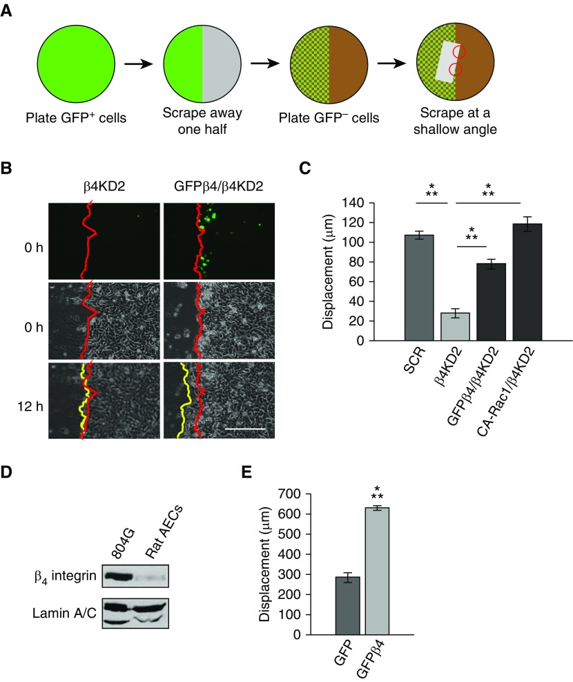
Figure 6.
Hybrid monolayers. (A) Schematic of procedure to create hybrid cell monolayers. A scrape (indicated by the gray rectangle) was made in a hybrid monolayer composed of β4KD and β4KD cells expressing GFP-tagged β4 integrin (GFPβ4) or CA-Rac1. The angle of the scrape allowed us to analyze healing of sheets of knockdown cells in which all follower cells were β4KD cells and the leading cells were β4KD cells (upper red circle in A) or either knockdown cells expressing GFPβ4 (GFPβ4/β4KD) or CA-Rac1 (CA-Rac1/β4KD) (lower red circle in A). (B) Fluorescence and phase–contrast images of the two different regions in A. Red lines indicate the edges of the original scratch, whereas the yellow lines indicate the wound edge at 12 hours after wounding. In the upper right panel, several GFPβ4 integrin–positive cells are close to the leading edge. Note that the GFPβ4/KD2 cell combination has begun to move over the wound, whereas the KD cells have not. Scale bar, 300 µm. (C) Quantification of hybrid sheet experiments (3 experiments, >10 scratches). ANOVA indicated differences between groups. Specific groups were compared by t test, as indicated. (D) Western blot of β4 integrin in rat bladder 804G cells and primary rat alveolar epithelial cells (AECs) shows low expression levels in freshly isolated cells. (E) Quantification of the leading-edge displacement of scratched epithelial sheets of rat AECs infected with adenovirus encoding GFP or GFPβ4 (17 scratches, t test). Results are presented as means (±SEM). ⁂P < 0.001.

