
Figure 6.
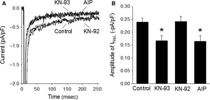
Decrease in IN aL in the presence of CaMKII inhibitors. Panel A, representative current traces recorded from four myocytes treated with no drug (control), KN‐93 (10 μmol/L), KN‐92 (10 μmol/L), and AIP (2 μmol/L), respectively. Panel B, summary of the results obtained from experiments shown in panel A. Bars represent the average amplitude of IN aL in control (n = 40/17) and in the presence of KN‐93 (n = 11/3), KN‐92 (n = 12/4), and AIP (n = 11/5). *P < 0.05 versus control.