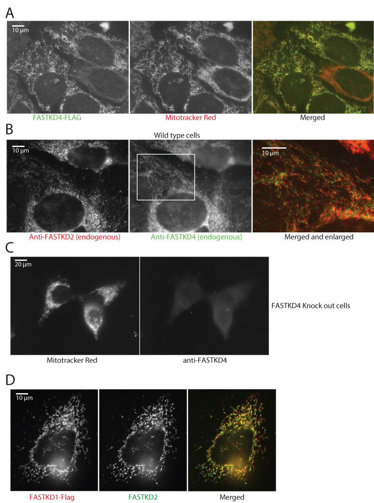
Figure 1.
Submitochondrial localization of FASTKD1 and FASTKD4. (A) FASTKD4 has a mitochondrial distribution, but is not concentrated in foci. 143B cells expressing Flag-tagged FASTKD4 were immunostained for FASTKD4 using an anti-Flag antibody (left) and with Mitotracker Red to reveal mitochondria (center). Right: merged image. Flag staining is in green, and Mitotracker Red is in red. (B) FASTKD4 shows a diffuse mitochondrial staining whereas FASTKD2 is present in discrete foci. Immunostaining of endogenous FASTKD2 (left) and endogenous FASTKD4 (center) in 143B cells using anti-FASTKD2 and FASTKD4 antibodies respectively. In the right panel, the images are merged (FASTKD4 in green and FASTKD2 in red) and enlarged (the enlarged area is indicated with a white rectangle in the center panel). (C) Specificity of the anti-FASTKD4 antibody. FASTKD4-KO cells were labeled with Mitotracker Red (left) and anti-FASTKD4 antibody (right). No specific FASTKD4 signal is visible in the FASTKD4-KO cells. (D) FASTKD1 localizes to MRGs. 143B cells were transfected with pCi FASTKD1-FLAG. Cells were then immunostained with antibodies against endogenous FASTKD2 to label MRGs (middle panels) or FLAG to label FASTKD1 (left panels). The merge (right panel) shows that FASTKD1 is in MRGs.

