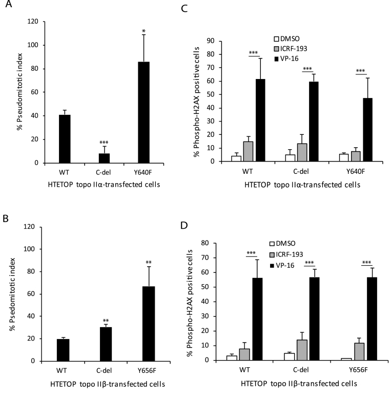
Figure 4.
Deletion of the CTD of topo IIα or topo IIβ or mutation of the homologous tyrosine Y640F in topo IIα or Y656F in topo IIβ dysregulates the decatenation checkpoint. (A) Pseudomitotic index of HTETOP cells expressing WT, C-del or Y640F mutant topo IIα. Columns represent the mean of three biological replicates and the bars indicate standard deviations. *P < 0.05, **P < 0.001 (two-sample t-test was used to compare WT topo IIα-transfected cells to C-del topo IIα-transfected cells or Y640F mutant topo IIα-transfected cells). (B) Pseudomitotic index of HTETOP cells expressing WT, C-del or Y656F mutant topo IIβ. Columns represent the mean of three biological replicates and the bars indicate standard deviations. **P < 0.01 (two-sample t-test was used to compare WT topo IIβ-transfected cells to C-del topo IIβ-transfected cells or Y640F mutant topo IIβ-transfected cells). (C) Determination of H2AX phosphorylation in HTETOP cells expressing WT, C-del or Y640F mutant topo IIα that were treated with DMSO, ICRF-193 or VP-16. Columns represent the mean of at least three biological replicates and the bars indicate standard deviations. ***P < 0.001 (two-way ANOVA, Student-Neuman-Keuls post-hoc test). (D) Determination of H2AX phosphorylation in HTETOP cells expressing WT, C-del or Y656F mutant topo IIβ that were treated with DMSO, ICRF-193 or VP-16. Columns represent the mean of at least three biological replicates and the bars indicate standard deviations. ***P < 0.001 (two-way ANOVA, Student-Neuman-Keuls post-hoc test).

