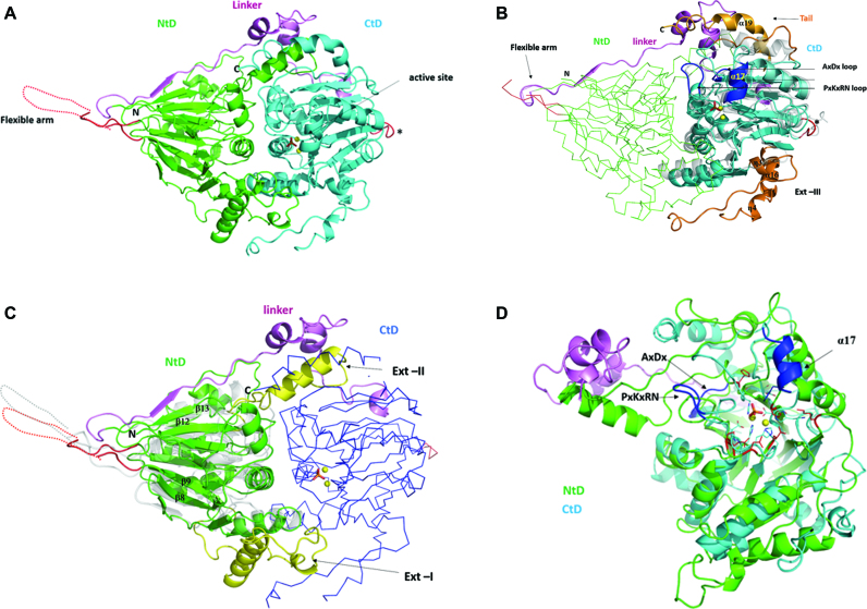
Figure 1.
Structure of Trz1. (A) Cartoon presentation of the Trz1 structure. The N-terminal domain (NTD) and C-terminal domain (CTD) are shown in green and cyan, respectively, and the linker is shown in purple. Two zinc ions are shown as yellow spheres and the phosphate ion is shown as sticks (present in the active site). The region corresponding to the unresolved flexible arm protruding from the NTD is shown as a dashed line and the loop replacing the flexible arm in the CTD is in red (labeled with an asterisk). (B) The CTD of Trz1 superimposed on BsuTrz (transparent gray, PDB code: 1y44). The core of the CTD is shown in cyan and the extensions are in orange. The conserved tRNA recognition elements α17 and loop PxKxRN and the nearby AxDx loop in the CTD are in blue. The NTD of Trz1 is shown as a green ribbon and the linker region is shown in purple. (C) The NTD of Trz1 superimposed on BsuTrz (transparent gray). The β-lactamase core of the NTD is shown in green and the extensions are in yellow. The CTD of Trz1 is shown as blue ribbon. (D) Superimposition of the NTD (green) and CTD (cyan) of Trz1 (linker in purple).

