Figure 3.
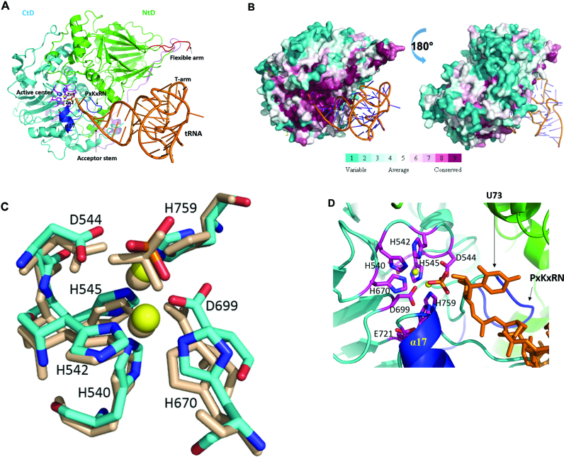
Model of Trz1 in complex with tRNA. (A) This model was generated by superimposition of Trz1 onto the BsuTrz/tRNA complex (PDB code: 2FK6). The N- and C-terminal halves and linker of Trz1 are shown in green/cyan and purple, respectively. For clarity, BsuTrz was omitted in the figure and only the tRNA (orange) is shown. Arrows indicate the position of the flexible arm and acceptor stem. The active center residues of Trz1 are shown as pink sticks, and zinc ions as yellow spheres. The α17 helix and the ‘PxKxRN’ and AxDx loops are shown in blue. (B) Sequence conservation projected onto the surface of Trz1 as generated with default parameters by the consurf webserver (http://consurf.tau.ac.il). The complex is shown in the same orientation as for panel A. (C) Zoom on the superimposition of the active site residues of Trz1 and BsuTrz. Only Trz1 residues are labeled and BsuTrz residues are shown as gray sticks. (D) Zoom on the active site of the Trz1/tRNA complex. The tRNA is shown as orange sticks and the uracil 73 is labeled.

