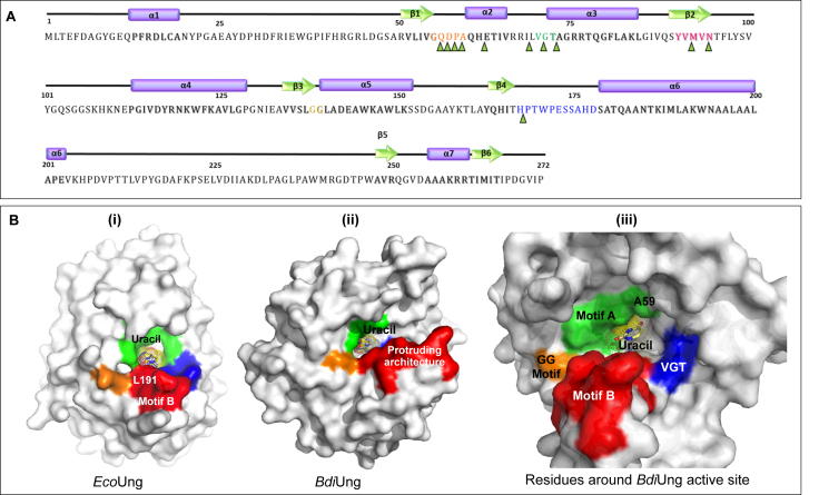
Figure 7.
Primary sequence and motifs of BdiUng. (A) Secondary structure elements and motifs are represented along with the primary sequence. Alpha helices are represented as purple cylinders while beta strands are shown as green arrows and corresponding regions in the primary sequence are shaded. Sequences are colored as follows: motif A: orange, PPS motif: green, VLLLN motif: pink, GS motif: yellowish green, sequence forming protruding structure (motif B): blue. Residues constituting ligand binding cavity are marked by green triangles. The nomenclatures of the motifs are done with respect to family 1 UDGs. (B) Surface diagrams of EcoUng (PDB: IEUI) and BdiUng generated using Pymol. (i) The conserved motifs in Ung are colored (motif A; green, motif PPS; blue, motif VLLLN; yellow, motif GS; orange and motif B; red). (ii) The corresponding regions of BdiUng to Ung have the same color. (iii) The residues surrounding the uracil binding pocket are shown in the surface diagram.

