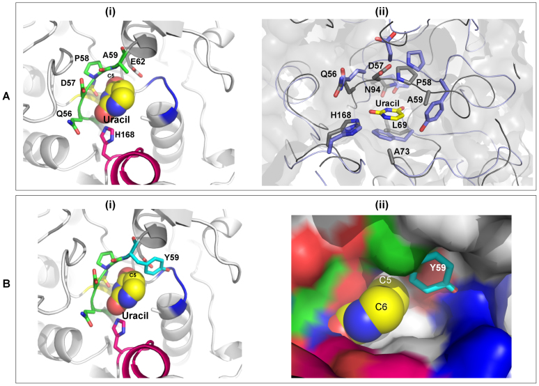
Figure 8.
Active site of BdiUng. (A) (i) The active site of BdiUng is shown along with uracil depicted as spheres. Motif A and H168 are highlighted. (ii) The ligand binding cavity of BdiUng and E. coli Ung (PDB Id: 1EUI) show high similarity. Overlap of BdiUng (grey) and E. coli Ung (blue) active site. The residues are labelled for BdiUng. (B) Model structures for mutation A59Y (i) BdiUng active site showing the A59Y mutation. (ii) Surface diagram of BdiUng showing the steric hindrance to substituents at C5 of uracil molecule caused by the A59Y mutation.

