
Figure 1:
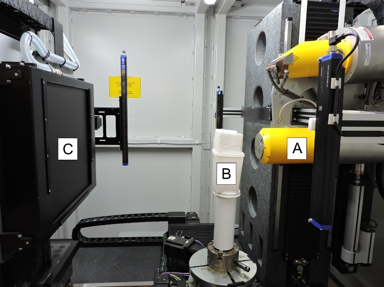
Photograph of the micro-CT scanner used during the study showing the fundamental components of the setup. A typical micro-CT scanner consists of an x-ray tube (A) that emits x-rays, which pass through a sample (B) before being recorded by an x-ray detector (C).