
Figure 4. IL-4 induces IL-7Rα expression and TSLP responsiveness of splenic DC's.
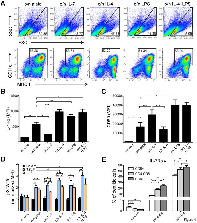
(A) Comparison of splenic DC populations after different o/n culturing conditions. (B-C) Statistical analysis of IL-7Rα (B) and CD80 (C) expression on splenic DC's, Bars indicate MFI (with +SEM). IL-7Rα and CD80 staining data is from three independent experiments (3 animals used in each experiment). (D) STAT5 phosphorylation in splenic DC's (either ex vivo or o/n cultured with indicated stimulations) in response to either vehicle (PBS,) or IL-7 or TSLP. The bars indicate normalized MFI of pSTAT5 (o/n cultured unstimulated sample was given the value 1). Data is from three independent experiments (three animals used in each experiment). (E) IL-7Rα upregulation in different DC subpopulations in response to IL-4 was measured. MFI and +SEM are indicated. *p<0.05, **p<0.01,***p<0.001, two-tailed, paired t-test was used.