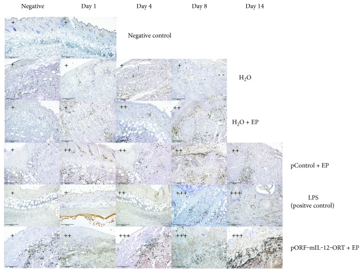
Figure 5.
Immunohistochemical staining of MHCII-positive cells of all the experimental groups (negative control, H2O, H2O + EP, LPS (positive control), pControl + EP, and pORF-mIL-12-ORT + EP) on day 1, day 4, day 8, and day 14 (11). A semiquantitative scoring system for immunopositive cells was used: (+) low, (++) moderate, and (+++) high positivity. The negative images of all groups are not related to a specific day. The images were taken under 20x magnification (numerical aperture 0.85).

