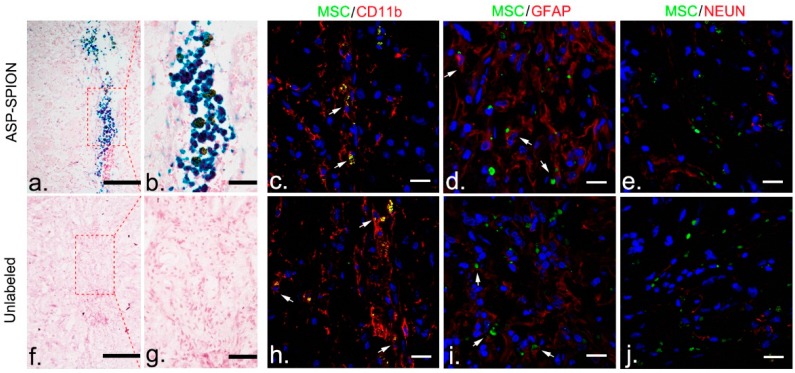
Figure 8.
Histopathology assessment of grafted cells. At 6 weeks after transplantation, Prussian blue staining micrographs show that blue-stained cells remained in the injection site, surrounded by extracellular positive aggregates, in animals treated with ASP-SPIONs-labeled cells (a,b). No positive iron-containing cells were found in animals grafted with unlabeled cells (f,g). Scale bar for a,b,c,f: 100 μm. Fluorescence immunostaining micrographs reveal that green fluorescent protein (GFP)-MSCs remained in the injection site in animals treated with ASP-SPIONs-labeled cells or unlabeled cells. Few GFP-cells were differentiated into GFAP positive astrocyte (d,i), but no cells into NeuN positive neurons (e,j). A small number of cells were phagocytized by macrophages (c,h). Scale bar for all immunostaining micrographs: 20 μm.

