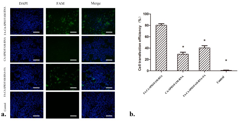
Figure 4.
In vitro cell transfection efficiency analysis. (a) Representative fluorescence microscopy micrographs show the uptake of siRNA in HepG2 cells after 30 min incubation with the superparamagnetic iron oxide nanoparticles (SPIO)-loaded cationic amylose complexed with FAM-labeled scramble siRNA (CA-SPIO/FAM-RNA), folate-conjugated CA-SPIO/FAM-RNA (FA-CA-SPIO/FAM-RNA), FA-CA-SPIO/FAM-RNA plus FA and untreated cells (control). Scale bar: 100 μm; and (b) flow cytometric data of cells incubated with FA-CA-SPIO/FAM-RNA, CA-SPIO/FAM-RNA, FA-CA-SPIO/ FAM- RNA plus FA and untreated cells (control). *: p < 0.05 compared with FA-CA-SPIO/FAM-RNA group.

