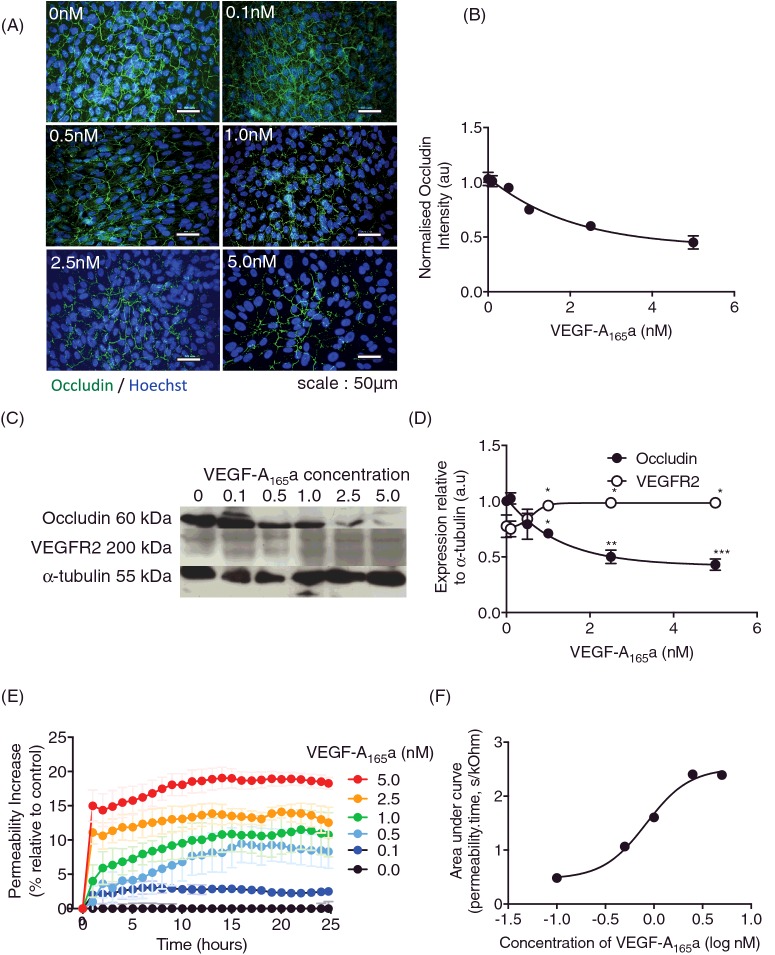
Figure 1. VEGF-A165a causes RPE tight junction breakdown.
RPE cells were treated with increasing doses of rhVEGF-A165a for 24 h (n=3, mean ± SEM). (A) Cells were fixed and stained for TJ marker, occludin (green) and nuclei co-stained with Hoechst (Blue). (B) Intensity was measured using ImageJ and normalized to untreated. (C) Protein lysate was extracted and immunoblotted for occludin and VEGFR2 expression (n=2, mean ± SD). Membranes were stripped and re-probed for α-tubulin expression to confirm equal loading of protein and densitometry (D). (E) RPE cells were plated on ECIS plates (n=3, mean ± SEM) to assess paracellular flux treated with a range of VEGF-A165a concentrations (0–5 nM). (F) Area under the curve was calculated over the 25-h period and plotted against concentration. The concentration that gave a 50% increase in permeability was determined by curve fitting using a four-parameter variable slope log(agonist) versus response curve in GraphPad Prism7; *p<0.05, **p<0.01, ***p<0.001, Figure A–C: one-way ANOVA with Dunnett's post-test; Figure E: two-way ANOVA with Bonferroni post-hoc, significance shown at 5-h intervals.

