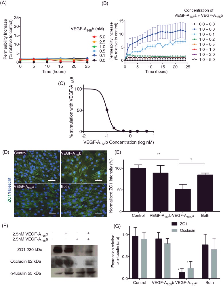
Figure 2. VEGF-A165b prevents VEGF-A165a -induced changes in tight junctions.
RPE cells were plated on ECIS plates (n=3, mean ± SEM) and treated with increasing concentrations (A) of VEGF-A165b (0–5 nM) or with different proportions (B) of VEGF-A165b: VEGF-A165a and TEER were measured over 24 h (n=3, mean ± SEM) and impedance was measured over 24 h; ***p<0.001, two-way ANOVA with Bonferroni post-hoc. (C) Inhibition of the RPE total solute flux induced by 1 nM VEGF-A165a calculated from the area under the curve; IC50=1 nM. Cells were stained (D) for ZO1 (green) and fluorescence intensity was calculated (E). Protein lysate was immunoblotted for ZO1 and occludin expression (F). Membranes were stripped and re-probed for α-tubulin expression and expression was quantified (G); n=3, one-way ANOVA, Tukey's post-hoc test, *p<0.05, **p<0.01.

