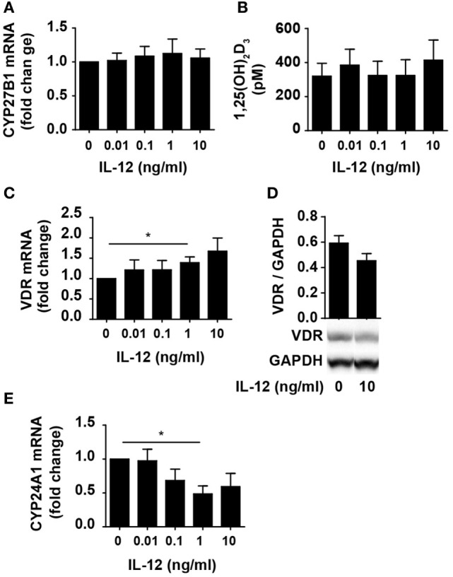
Figure 2.

IL-12 does not inhibit 1,25(OH)2D3 production or VDR expression and function in CD4+ T cells. Relative CYP27B1 (A), VDR (C), and CYP24A1 (E) expression in T cells activated for 72 h in the presence of 100 nM 25(OH)D3 and the indicated concentration of IL-12. Data are normalized to activated T cells incubated with 100 nM 25(OH)D3 in the absence of IL-12 (mean + SEM, n ≥ 6). (B) 1,25(OH)2D3 production in T cells activated for 72 h in the presence of 100 nM 25(OH)D3 and the indicated concentration of IL-12 (mean + SEM, n ≥ 4). (D) Representative Western blot (lower panel) and quantification (upper panel) of VDR with GAPDH as loading control from T cells activated for 72 h in the presence of 100 nM 25(OH)D3 and in the presence or absence of 10 ng/ml IL-12 (mean + SEM, n = 4). Western blots including protein ladder are shown in the Figure S1 in Supplementary Material.
