Fig. 5.

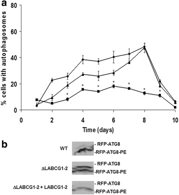
Autophagosome formation in promastigotes of L. major lines. a The proportion of promastigotes from different lines including: control (WT) (diamonds), ΔLABCG1-2 (squares), and ΔLABCG1-2 + LABCG1-2 (triangles) expressing RFP-ATG8 was assessed during the growth curve in vitro. Data are the mean ± SD from three independent experiments. Statistical differences relative to the WT values were determined using the Student’s t test (*P < 0.05). b Western blot analysis of RFP-ATG8 in different Leishmania lines. Promastigote cell extracts from different lines (WT, ΔLABCG1-2 and ΔLABCG1-2 + LABCG1-2) transfected with RFP-ATG8 were collected during the stationary growth phase, separated by SDS-PAGE in the presence of 6 M urea and analysed by Western blot. A Western blot assay representative of at least three independent experiments is shown
