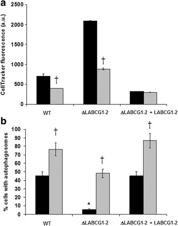
Fig. 7.

Relationship between thiol levels and autophagosome formation in L. major lines. a Parasites (WT, ΔLABCG1-2 and ΔLABCG1-2 + LABCG1-2) transfected with pNUS RFP-ATG8 were pre-incubated for 48 h in M-199 without (black histograms) or with (grey histograms) 3 mM BSO in order to deplete thiol levels. After 8 days in culture, promastigotes (4 × 10⁶) were collected and incubated with 2 μM CellTracker™ for 15 min. Fluorescence intensities were measured by flow cytometry. b In parallel, we measured the proportion of promastigotes expressing RFP-ATG8 with (grey histograms) or without (black histograms) BSO treatment by counting autophagosomes using a fluorescence microscope. Data are the mean ± SD from three independent experiments. Significant differences were determined by the Student’s t-test (*P < 0.01 versus WT; †P < 0.01 versus non-treated parasites)
