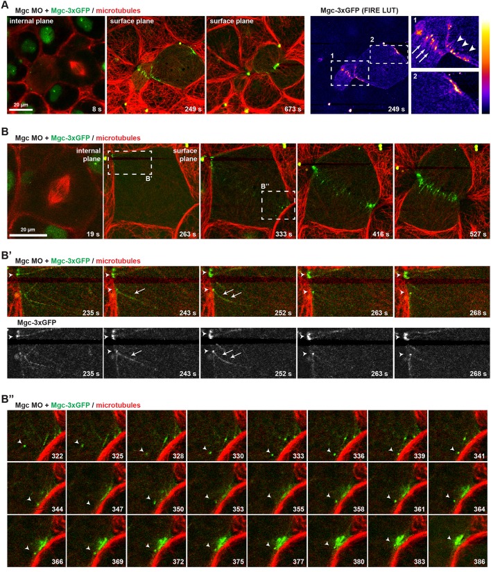
Fig. 1.
MgcRacGAP localizes to microtubule plus ends at the equatorial cortex as cytokinesis initiates. (A) Still images from a single z-plane time-lapse movie of a X. laevis embryo co-injected with Mgc MO+Mgc–3×GFP and a probe for MTs (2×mChe–EMTB). Mgc–3×GFP (green) localizes in the nucleus of interphase cells, at overlapping central spindle MTs, as well as at individual MTs at the equatorial cortex prior to furrowing. A FIRE look-up table (LUT) plugin was applied to the Mgc–3×GFP channel to highlight Mgc localization, and enlarged regions are shown on the right (arrows, overlapping central spindle MTs; arrowheads, individual MTs at equatorial cortex). (B) Still images from a single z-plane time-lapse movie of a X. laevis embryo co-injected with Mgc MO+Mgc–3×GFP and 2×mChe–EMTB. The dashed boxes in B indicate regions that are enlarged in B′ and B″. The frames in B′ show Mgc–3×GFP decorating equatorial astral MTs (arrow) and Mgc–3×GFP puncta at MT plus ends (arrowheads). The frames in B″ show directed movement of an Mgc–3×GFP puncta (arrowheads), apparently along astral MTs, as Mgc–3×GFP forms clusters at the equatorial cell cortex.

