Fig. 1.
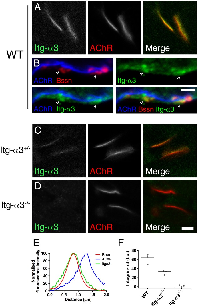
Localisation of integrin-α3 at the AZs of NMJs. Immunofluorescence staining for integrin-α3 (Itg-α3) in E18.5 sternomastoid muscles from wild type (WT) (A), integrin-α3+/− (C) and integrin-α3−/− (D) mice. Fluorescently conjugated α-bungarotoxin was used to mark postsynaptic acetylcholine receptors (AChRs). (B) Co-labelling of integrin-α3 with presynaptic AZ marker, bassoon (Bssn), in WT E18.5 muscles. (E) Line scans across the synaptic interface, showing colocalisation of bassoon and integrin-α3, and their spatial separation with postsynaptic AChRs. (F) Fluorescence intensity of integrin-α3 staining in each genotype. Single confocal slices (A,C,D); z-stack of three confocal slices 0.4 μm apart, at high (×63) magnification (B). (E) Averaged traces from ten AZs across 4 WT NMJs. (F) Ten WT, 18 integrin-α3+/− and 8 integrin-α3−/− NMJs, taken from three animals/genotype (data points for each animal plotted with median indicated by a line). Scale bars: 5 μm (A,C,D); 2 μm (B).

