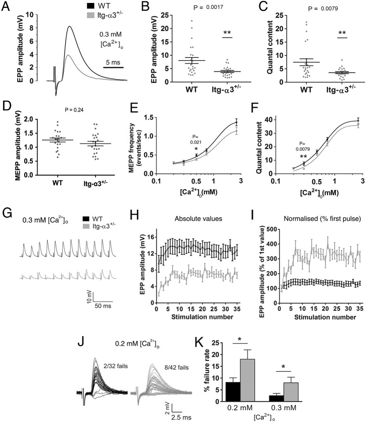
Fig. 5.
Impaired synaptic vesicle release in integrin-α3+/− NMJs at reduced external Ca2+ concentrations. Ex vivo electrophysiology on adult (5 months) phrenic nerve and diaphragm preparations, with external buffer containing 0.3 mM Ca2+. EPP amplitude (A,B) and quantal content (C) were significantly reduced in integrin-α3+/− NMJs compared to in WT. No difference in MEPP amplitude was observed, indicating a normal postsynaptic response (D). Two indicators of vesicle release probability, MEPP frequency (E) and quantal content (F) were plotted against varying concentrations of Ca2+. Dose–response curves for integrin-α3+/− NMJs are significantly shifted to the right, indicating a very slightly reduced sensitivity of nerve terminals to Ca2+. 50 Hz repetitive stimulation, at 0.3 mM Ca2+ (G-I). Traces were averaged and plotted as absolute EPP amplitude values (H), or normalised to the first EPP value in the train (I). Normalised traces revealed an enhancement in short-term facilitation over the first few stimuli in integrin-α3+/− NMJs over that of WT (I). Representative 1 Hz traces at 0.2 mM Ca2+ (J). Increased rate of neurotransmission failure in integrin-α3+/− endplates at 0.2 mM and 0.3 mM Ca2+ (K). All recordings are from 3-6 animals/genotype; n=22-24 recordings, except in F-H, where n=12-15 recordings. All graphs are mean± s.e.m., Student's t-test; two-way ANOVA with mixed model for panels E and F.

