
Fig. 7.
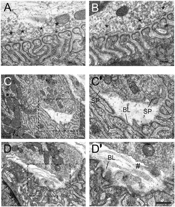
Nerve terminal detachment in integrin-α3+/− NMJs. Electron micrographs of diaphragm NMJs from 2-month-old WT (A) and integrin-α3+/− (B-D′) mice. NMJs in integrin-α3+/− mice resembled those of WT in their ultrastructure: AZs (asterisks), postsynaptic folds and alignment of AZs with folds; full quantification of these parameters is shown in Table 1. In integrin-α3+/− mice, instances of nerve terminal detachment were observed (C-D′), comprising 13.0% of all boutons analysed, with none in WT. C′ and D′ are enlarged images of the regions indicated in C and D, respectively. Schwann cell processes (SP) were sometimes observed to encase parts of the detached nerve terminals. Basal lamina (BL) remains largely intact on the postsynaptic muscle side. Regions of detached nerve terminals were sometimes left exposed and without Schwann cell protrusions (marked with #). n=56 synaptic boutons in WT, and 63 in integrin-α3+/− mice, across three animals per genotype. Scale bar: 500 nm, except for C and D, where the same bar represents 1 µm.