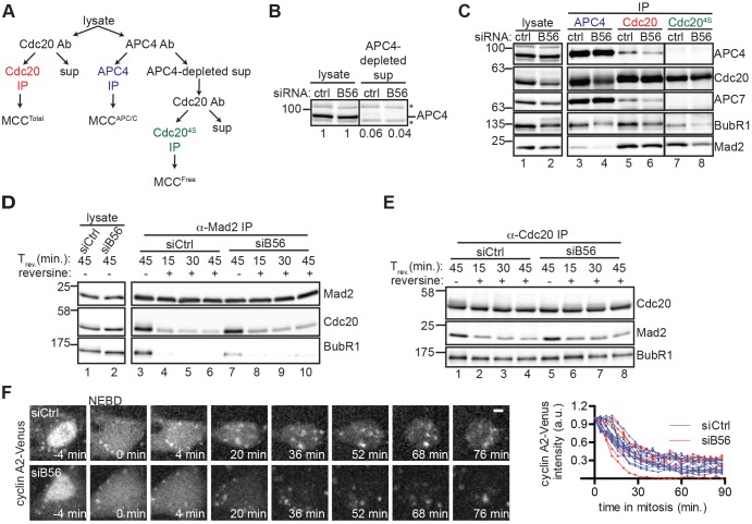
Fig. 2.
B56 depletion does not increase the amount or stability of the mitotic checkpoint complex. (A) Schematic of experiment to isolate different pools of MCC from whole-cell lysates, including total MCC (Cdc20 IP), MCC bound to APC/C (co-precipitation in APC4 IP), and MCC free of APC4 [isolated by subjecting APC4-depleted supernatant (4S) to Cdc20 IP (Cdc204S IP)]. (B) Comparison of APC4 levels in siCtrl and siB56 whole-cell lysates versus APC4-depleted supernatant by quantitative western blotting. Numbers indicate the relative amount of APC4 in lysate versus APC4-depleted supernatant. *, non-specific band. (C) Whole-cell lysate (lanes 1 and 2) and IPs performed as outlined in A (lanes 3-8) were probed by western blotting. In B,C, solid vertical lines indicate that intervening lanes have been removed. (D,E) MCC is not stabilized in siB56 cells. Mitotic HeLa siCtrl and siB56 cells were incubated with MG132 and reversine or DMSO before lysis and then analyzed directly (D, lanes 1,2) or split for immunoprecipitation of Mad2 (D, lanes 3-10) and Cdc20 (E) and probed by western blotting. (F) Cyclin A proteolysis is not delayed in siB56 cells. RPE1 CCNA2Venus/+ siCtrl or siB56 cells in nocodazole were imaged live. (Left) Micrographs of Venus (cyclin A2) are shown with time (min) relative to nuclear envelope breakdown (NEBD). (Right) The fraction of cyclin-A2–Venus fluorescence remaining as a function of time in mitosis is plotted. Each line indicates a single cell. Scale bar: 5 μm. Results in B–F are representative of three independent experiments. Ab, antibody; sup, supernatant; Trev, time before or after reversine addition.

