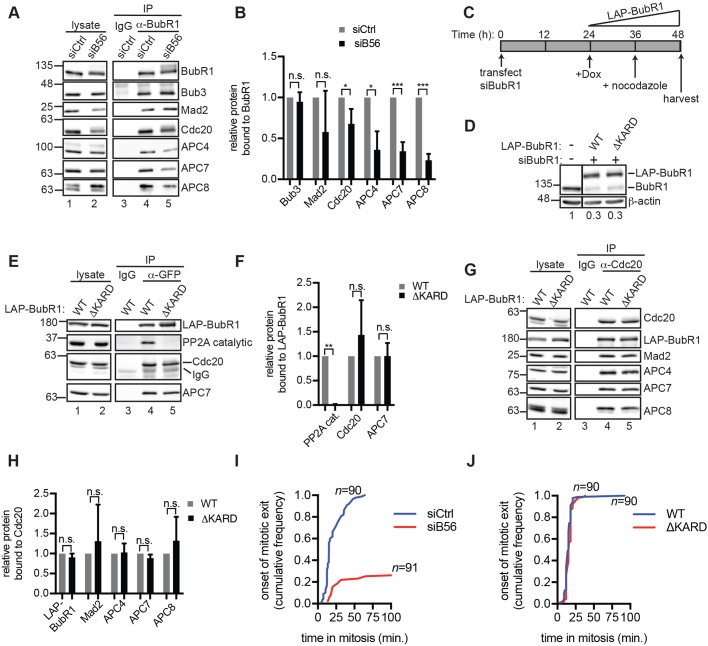
Fig. 4.
PP2AB56-dependent stimulation of APC/CCdc20 assembly does not require direct binding between B56 and BubR1. (A) BubR1 association with APC/C is reduced in siB56 cells. Mitotic siB56 or siCtrl HeLa cells were used for BubR1 (lanes 4,5) or control (lane 3) IgG IPs and analyzed by western blotting. (B) Quantification of three replicate experiments as in (A). (C–H) Deletion of the KARD in BubR1 does not alter APC/C–Cdc20 association. (C) Schematic for BubR1 depletion and rescue in HeLa cells. (D) BubR1-rescue lysates were analyzed alongside lysates from untreated cells by quantitative western blotting. Endogenous BubR1 signals were normalized to the actin signal, and the value in the untreated sample was set to 1. Solid line indicates intervening lanes have been cropped. (E–H) Lysates (lanes 1,2) and IPs (lanes 3-5) of GFP (E) or Cdc20 (G) were analyzed by western blotting. The experiments in E,G were performed three times and the quantifications are shown in F and H, respectively. (I,J) Comparison of mitotic exit delay in siB56 cells versus BubR1-rescue cells. siCtrl and siB56 cells (I) or BubR1-rescue cells (J) were incubated in nocodazole and reversine and imaged live. Plotted is the fraction of cells that exited mitosis as a function of time after mitotic entry. n, total number of cells analyzed from three independent experiments. Bars are mean±s.d. n.s., not significant (P>0.05); *P<0.05, **P<0.005, ***P<0.0005, Student's two-tailed t-test.

