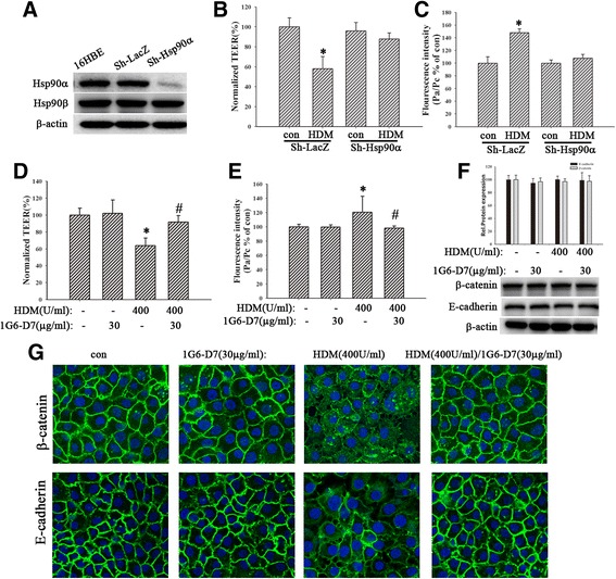
Fig. 3.

Both silencing of Hsp90α and the eHsp90α mAb, 1G6-D7, restored HDM-induced epithelial barrier dysfunction. a The expression of Hsp90α in sh-Hsp90α group decreased remarkably. b and c Sh-Hsp90α cells were treated with HDM (400 U/ml) for 24 h, then TEER values and FITC-DX were measured immediately. d and e Cells were pretreated with 1G6-D7 (30 μg/ml) for 2 h and then treated with HDM (400 U/ml) for 24 h. TEER values and FITC-DX were measured. fThe expression of E-cadherin and β-catenin proteins was detected by western blotting analysis. g Immunofluorescence staining was used to show the localization of E-cadherin or β-catenin. Data are mean ± SD of four independent experiments.* P < 0.05 versus con group; # P < 0.05 versus HDM group
