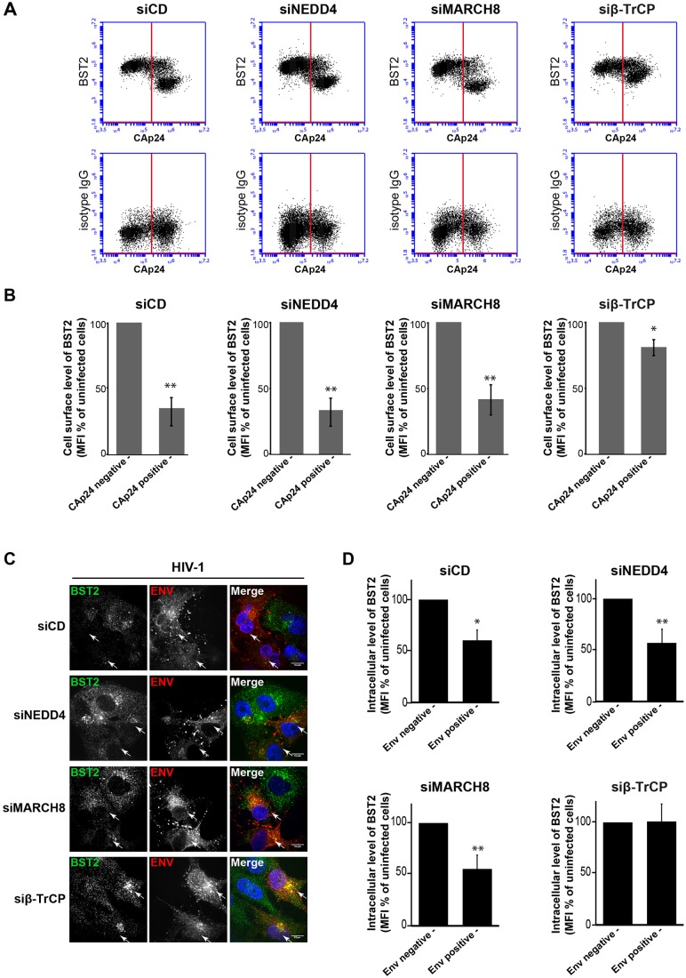
Fig. 7.
NEDD4 and MARCH8 are not required for Vpu-induced BST2 downregulation. (A,B) Effects of NEDD4, MARCH8 or β-TrCP silencing on Vpu-induced cell surface downregulation of BST2. HeLa cells that had been transfected with the indicated siRNA were infected with VSV-G-pseudotyped HIV-1 NL4-3 WT. Twenty-four hours later, cells were surface-stained for BST2 or an isotype IgG control. The cells were then fixed, permeabilized and stained for Gag using a FITC-conjugated monoclonal antibody against CAp24. The cells were then processed for flow cytometry analysis. (A) Dot plot. Vertical lines indicate the gates set using non-infected cells stained as indicated. Left gate, non-infected cells; right gate, infected cells. (B) Bar graph representation of the cell surface level of BST2 in CAp24-negative cells (left bars) and CAp24-positive cells (right bars) for each siRNA condition. Values are expressed as the mean fluorescence intensity (MFI) for BST2 staining minus those of the isotype control, normalized to those of non-infected cells set as 100%. Bars represent the mean±s.d. (n=3); **P<0.01, *P<0.05 (Student's t-test). (C,D) β-TrCP is required for Vpu-induced BST2 degradation. (C) Infected siRNA-treated HeLa cells were permeabilized before fixation, and intracellular BST2 (green) and HIV Env (red) were labeled with specific antibodies. Nuclei were stained with DAPI. Cells were imaged by confocal microscopy. Env staining discriminates infected cells (arrows) and non-infected cells. Scale bars: 10 µm. Images are representative of three independent experiments. (D) Infected siRNA-treated cells were fixed, permeabilized and stained for intracellular BST2 and HIV-1 Env using specific antibodies before analysis by flow cytometry. BST2 expression levels on infected and non-infected cells, respectively, were expressed as in B. Bars represent the mean±s.d. (n=3);**P<0.01, *P<0.05 (Student's t-test).

