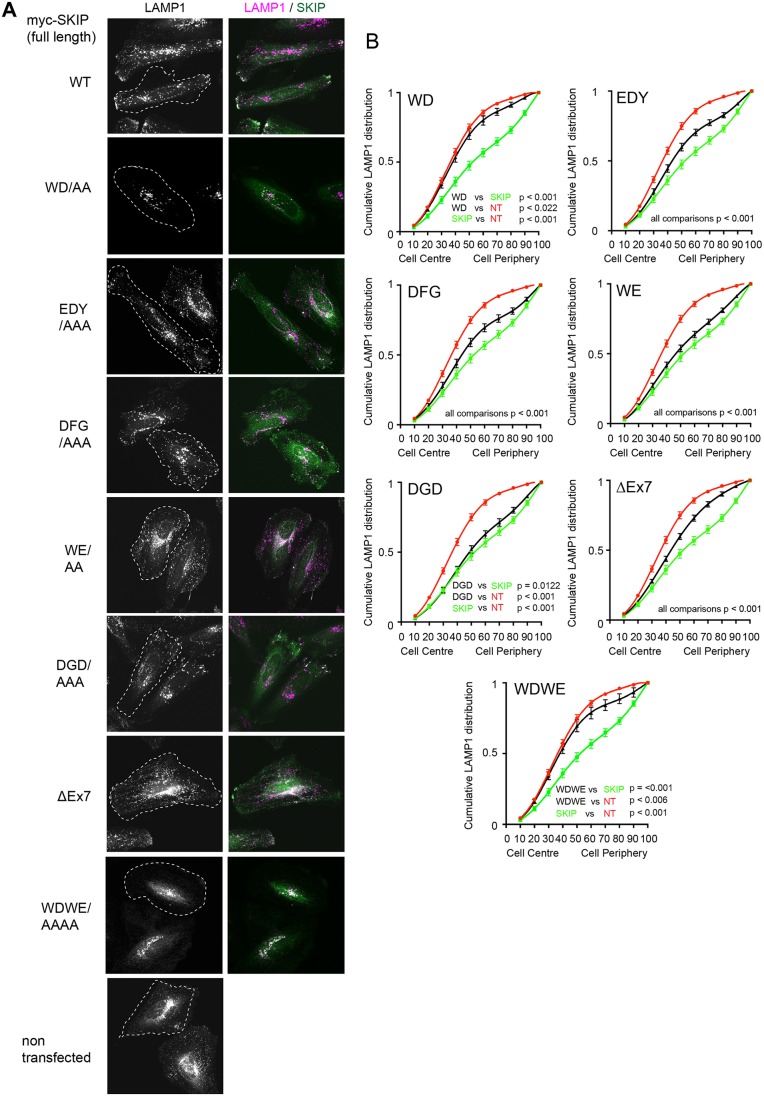
Fig. 7.
Specific disruption of SKIP–KHC binding limits the capacity of SKIP to promote the peripheral dispersion of lysosomes. (A) Immunofluoresence images showing late endosomes and lysosomes (LAMP1, magenta) in HeLa cells transfected with full length Myc–SKIP (green) and the indicated KHC-binding mutants. (B) Graphs showing quantification of lysosome distribution from 45 cells in 3 replicates. Error bars show ±s.e.m. Non-transfected (NT, red) and wild-type Myc–SKIP-transfected (green) curves are reproduced across all graphs. Black line indicates the distribution of lysosomes in cells transfected with the indicated mutant. Statistical analysis of pairwise comparison of curves was carried out as described in the Materials and Methods.

