Fig. 3.

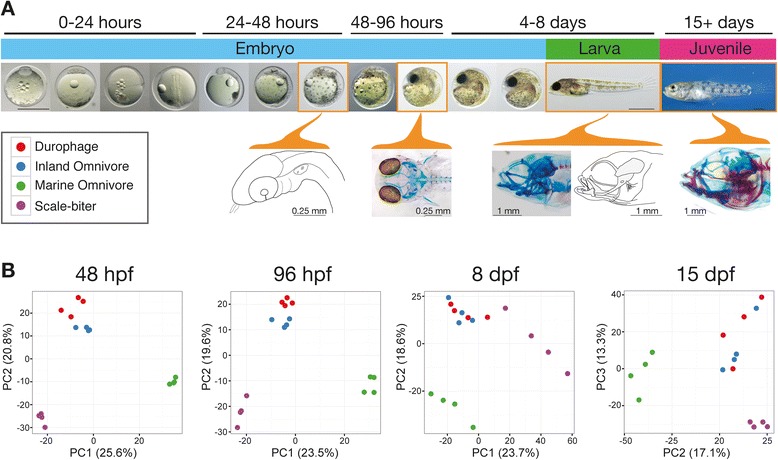
Gene expression patterns differ among species at all sampled stages. a Overview of pupfish development. The four developmental stages sampled in the current study are outlined with orange boxes. Camera lucida drawings and photos of fish stained for cartilage (blue) and bone (red) show head morphology at each of the sampled stages. At 48 hpf, fish resemble the pharyngula stage of zebrafish with migratory neural crest cells aggregated in the undifferentiated pharyngeal arches. By 96 hpf, the neurocranium and jaws first stain positive for cartilage (blue). Hatching 8 dpf larval fish have a mostly cartilaginous skull, but note the early ossification of dermal jaw bones that are highlighted in the camera lucida drawing. Morphological differences among pupfish jaws can be first measured in 15 dpf juvenile fish during a period of growth and increased bone deposition. b Species are separated by gene expression patterns along the first 2–3 principal component axes indicating a major effect of species in our dataset. A single PC axis typically separates the marine population from all three San Salvador taxa mirroring phylogenetic relationships from Fig. 2, while the inland omnivore samples tend to be more similar to the durophage samples than to samples from the morphologically similar marine omnivore population
