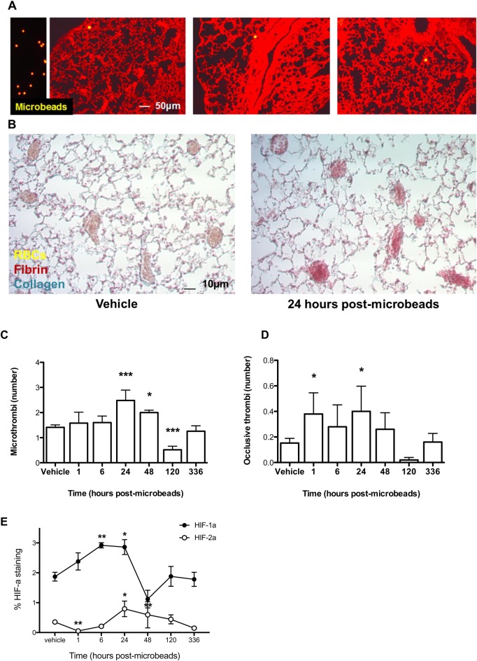
Fig. 1.
Administration of intravenous microbeads induces pulmonary microthrombosis. (A) Microbeads (yellow, 15 μm diameter) either isolated (left panel) or seeded intravenously into the pulmonary microvasculature at day 14 post-administration. (B) Representative lung sections were stained with MSB at day 1 post administration of intravenous microbeads or vehicle. (C) Number of total and (D) occlusive pulmonary microthrombi at various times following intravenous administration of microbeads or vehicle. (E) Pulmonary HIF1α and HIF2α levels at various times post-administration of intravenous microbeads. N=5/group. RBCs, red blood cells. *P<0.05 and ***P<0.001 versus vehicle-treated controls; unpaired two-tailed Student t-tests. Means±s.e.

