Abstract
Background
We previously reported that lifetime consumption of soy proteins or whey proteins reduced the incidence of azoxymethane (AOM)-induced colon tumors in rats. To obtain insights into these effects, global gene expression profiles of colons from rats with lifetime ingestion of casein (CAS, control diet), soy protein isolate (SPI), and whey protein hydrolysate (WPH) diets were determined.
Results
Male Sprague Dawley rats, fed one of the three purified diets, were studied at 40 weeks after AOM injection and when tumors had developed in some animals of each group. Total RNA, purified from non-tumor tissue within the proximal half of each colon, was used to prepare biotinylated probes, which were hybridized to Affymetrix RG_U34A rat microarrays containing probes sets for 8799 rat genes. Microarray data were analyzed using DMT (Affymetrix), SAM (Stanford) and pair-wise comparisons. Differentially expressed genes (SPI and/or WPH vs. CAS) were found. We identified 31 induced and 49 repressed genes in the proximal colons of the SPI-fed group and 44 induced and 119 repressed genes in the proximal colons of the WPH-fed group, relative to CAS. Hierarchical clustering identified the co-induction or co-repression of multiple genes by SPI and WPH. The differential expression of I-FABP (2.92-, 3.97-fold down-regulated in SPI and WPH fed rats; P = 0.023, P = 0.01, respectively), cyclin D1 (1.61-, 2.42-fold down-regulated in SPI and WPH fed rats; P = 0.033, P = 0.001, respectively), and the c-neu proto-oncogene (2.46-, 4.10-fold down-regulated in SPI and WPH fed rats; P < 0.001, P < 0.001, respectively) mRNAs were confirmed by real-time quantitative RT-PCR. SPI and WPH affected colonic neuro-endocrine gene expression: peptide YY (PYY) and glucagon mRNAs were down-regulated in WPH fed rats, whereas somatostatin mRNA and corresponding circulating protein levels, were enhanced by SPI and WPH.
Conclusions
The identification of transcripts co- or differentially-regulated by SPI and WPH diets suggests common as well as unique anti-tumorigenesis mechanisms of action which may involve growth factor, neuroendocrine and immune system genes. SPI and WPH induction of somatostatin, a known anti-proliferative agent for colon cancer cells, would inhibit tumorigenesis.
Keywords: colon cancer, soy, whey, gene expression profiling, neuro-endocrine, microarray, rat
Background
Colorectal cancer (CRC) is the third most common cancer and the third leading cause of cancer-related mortality in the U.S. [1,2]. Estimated new cases of colon cancer were 79,650 for men and 73,530 for women in 2004 [1]; approximately $6.3 billion is spent in the United States each year on treatment of CRC [2]. Accumulating evidence suggests that diet is an important environmental factor in the etiology of CRC. High consumption of red meats, animal fats, chocolate, alcohol and refined cereals are linked to higher incidence of these cancers in Western societies [3-5], whereas protective effects of fruits, vegetables and whole grains have been suggested [5].
Soy foods and soybean constituents have received considerable attention for their potential role in reducing cancer risk [6,7]. Our laboratories reported the protective effects of lifetime ingestion of soy protein isolate (SPI) on azoxymethane (AOM)-induced colon cancer in rats [8]. Similarly, the effect of whey protein hydrolysate (WPH) in the diet to reduce colon tumor incidence has been reported by us and others [9-11]. Several hypotheses have been proposed to account for soy and whey protein-induced anti-tumorigenesis. For example, soy isoflavones have been proposed to play a key role in soy's anti-cancer functions [12]. Yanagihara et al., among others, reported that genistein inhibits colon cancer cell proliferation and stimulates apoptosis in vitro [13-15]. However, subcutaneous administration of genistein to mice did not confirm these in vitro effects [16]. Holly et al. reported that soy sphingolipids inhibit colonic cell proliferation, and suggested that this may partially account for its anticancer benefits [17]. Other reports indicate that soy diets inhibit tumorigenesis by regulating the synthesis or activities of specific proteins. For example, Rowlands et al. reported that dietary soy and whey proteins down-regulate expression of liver and mammary gland phase I enzymes involved in carcinogen activation [18]. Elevated activities of phase II detoxification enzymes were reported in soy-fed rats [19,20]. Such dietary effects may result in lower tissue concentrations of activated carcinogen. The anticancer properties of whey proteins have been ascribed to their ability to elevate cellular levels of the antioxidant glutathione [21,22]. Moreover, the whey protein, α-lactalbumin, inhibits proliferation of mammary epithelial cells in vitro [11]. The anticancer properties of whey may also relate to its immune system-enhancing actions [23].
Despite extensive research, there is no consensus for anti-cancer mechanism(s) of soy and whey, which will undoubtedly involve multiple interrelated processes, pathways and many components. Many of the same molecular and biochemical changes underlying human colon cancer are observed in the azoxymethane (AOM)-induced rat colon cancer model [24]. Moreover, previous studies suggest a different molecular etiology for tumors of the proximal and distal colon in this model and in human colon [24,25]. Differential dietary effects on proximal vs. distal colon DNA damage were noted [26] and Westernization of the human diet is thought to have favored a shift of tumors from distal to more proximal locations [27]. Thus, region-specific localization of dietary effects on colon tumorigenesis is an important factor to consider in any molecular analysis of CRC. Here, we use Affymetrix high-density oligonucleotide microarrays to determine the expression profiles of non-tumor (i.e., normal) tissue in proximal colons (PC) of rats, subjected to lifetime diets containing casein (CAS, control diet), soy protein isolate (SPI), or whey protein hydrolysate (WPH) and which were administered AOM to induce tumors. We hypothesized that genes whose expression contributes to anti-tumorigenesis would be regulated in parallel by SPI and WPH; in addition, changes unique to each diet might also be apparent.
Results
Validation of the microarray approach
Quality control steps ensured that the RNA used for microarray and real-time RT-PCR analysis was of high quality. These steps included evaluation of the RNA with the RNA 6000 Nano Assay and assessment of the cRNA hybridization to GeneChips by comparison of data obtained for probe sets representative of 5' and 3' ends of control genes. All RNA samples had an A260/280 absorbance ratio between 1.9 and 2.1. The ratio of 28S to 18S rRNA was very close to 2 on RNA electropherograms, and signal ratios below 3 were noted for 3' vs. 5' probe sets for β-actin and glyceraldehyde-3-phosphate dehydrogenase (per Affymetrix user guidelines) after hybridization.
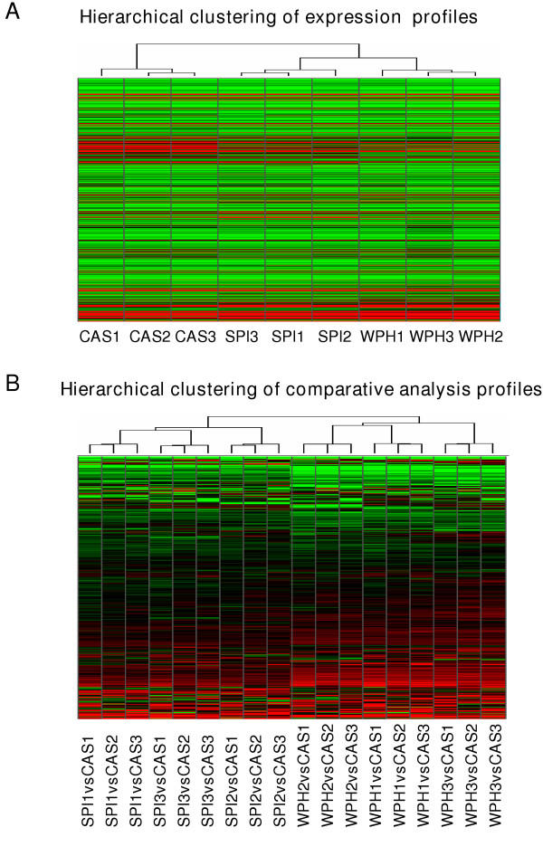
Total false change rates (TFC) were determined following Affymetrix-recommended guidelines [28], except that the inter-chip comparisons used cRNA targets made in parallel starting from the same RNA pool. Inter-chip variability, measured as TFC%, was 0.25% – 0.6% and well below the suggested 2% cutoff (Table 1). These values confirmed the fidelity and reproducibility of the microarray procedures used. Unsupervised nearest-neighbor hierarchical clustering identified differences in proximal colon gene expression profiles of CAS, SPI and WPH groups (Figs. 1 and 2), indicating that the type of dietary protein has a major effect on gene expression in normal proximal colon tissue of AOM-treated rats. Interestingly, the overall gene expression profiles for SPI and WPH groups were more similar to each other than each was to the CAS group (Fig. 1A).
Table 1.
Inter-chip variability
| Diet group | Number of arrays | TFC (%)* |
| CAS | 3 | 0.252 ± 0.138 |
| WPH | 3 | 0.369 ± 0.025 |
| SPI | 3 | 0.570 ± 0.165 |
*TFC (Total false change) = false change rate (decreased category) + false change rate (increased category), as described in ref. 28; TFC reported as mean ± SEM, TFC should be no more than 2% (Affymetrix).
Figure 1.
Hierarchical clustering of proximal colon gene expression profiles. A. Clustering of nine PC global gene expression profiles (8799 genes); n = 3 profiles each for CAS, SPI and WPH. Each cell represents the expression level of an individual gene in each sample (green = low expression, black = middle expression, red = high expression). The dendrogram reflects the extent of relatedness of different profiles; the shorter branch-point of the SPI and WPH trees indicates the greater similarity between these profiles. B. Clustering of 18 global comparative expression profiles including 9 of SPI vs. CAS and 9 of WPH vs. CAS profiles. Each row in the heat map represents the relative expression level of a given gene across all comparisons (red = up regulated, black = unchanged, green = down regulated).
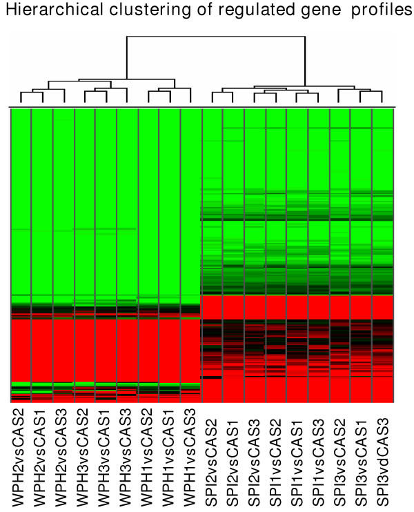
Figure 2.
Hierarchical clustering of 211 differentially expressed genes in either SPI or WPH. The differential expression data are taken only from the pairwise comparison analysis, with CAS profiles used as baseline. Each cell in the heat map represents the relative expression level of a given gene in an individual comparison analysis (red = up regulated, black = unchanged, green = down regulated). The dendrogram reflects the relatedness of different profiles.
Differentially expressed genes
Multiple filtering criteria were applied to the microarray data set so as to identify differentially expressed colon transcripts in rats fed SPI, WPH or CAS; results are reported only for transcripts that passed all three analytical filters used: DMT t-test, SAM and pair-wise comparison survival methods. Among the 8799 genes and ESTs examined with the rat U34A array, we identified 31 induced and 49 repressed genes in proximal colons of SPI-fed rats, whereas 44 induced and 119 repressed genes were detected in WPH-fed rats (Tables 2, 3, 4, 5). Interestingly, more down- than up-regulated genes were noted for both SPI and WPH. Additionally, 37 genes were co-repressed, whereas only two were co-induced by SPI and WPH (Table 6). More than 90% of identified genes in WPH and SPI animals showed the same direction of change relative to CAS. This is visually apparent in the hierarchical clustering output (Fig. 2).
Table 2.
Down-regulated genes in rats fed with WPH diet*
| Category and Gene Name | Probe Set GB Accession No. | Fold Change | P value |
| Cell adhesion | |||
| Embigin | AJ009698 | -6.57 | 0 |
| Cadherin 17 | L46874 | -4.8 | 0.036 |
| Cadherin | X78997 | -3.36 | 0.004 |
| Protein tyrosine phosphatase | M60103 | -2.64 | 0.004 |
| Cytokeratin-8 | S76054 | -2.71 | 0 |
| Trans-Golgi network integral membrane protein TGN38 | X53565 | -4.92 | 0.012 |
| Tumor-associated calcium signal transducer 1 | AJ001044 | -9.37 | 0.001 |
| Claudin-3 | AJ011656 | -7.55 | 0.02 |
| Claudin-9 | AJ011811 | -5.12 | 0 |
| Cell cycle/growth control | |||
| Mapk6 | M64301 | -2.61 | 0.003 |
| Epithelial membrane protein 1 | Z54212 | -4.67 | 0.015 |
| Glucagon | K02813 | -7.73 | 0.005 |
| Peptide tyrosine-tyrosine (YY) | M17523 | -4.56 | 0.001 |
| Src related tyrosine kinase | U09583 | -3.31 | 0.033 |
| FGF receptor activating protein | U57715 | -4.25 | 0.002 |
| Cyclin D1 | D14014 | -1.97 | 0.001 |
| Neu oncogene | X03362 | -2.61 | 0.017 |
| Defense/immunity protein | |||
| Seminal vesicle secretion protein iv | J00791 | -5.35 | 0.001 |
| Putative cell surface antigen | U89744 | -5.22 | 0.008 |
| Decay accelerating factor GPI | AF039583 | -6.12 | 0 |
| Beta defensin-1 | AF093536 | -26.78 | 0.001 |
| Detoxification/antioxidation | |||
| Glutathione S-transferase | J02810 | -5.17 | 0 |
| Glutathione S-transferase Yb | X04229 | -9.33 | 0 |
| J03914 | -2.43 | 0.002 | |
| Glutathione S-transferase, alpha 1 | K01932 | -3.07 | 0.002 |
| Glutathione transferase, subunit 8 | X62660 | -6.42 | 0.001 |
| Glutathione S-transferase Yc1 | S72505 | -3.69 | 0.004 |
| Glutathione S-transferase Yc2 | S72506 | -21.38 | 0.008 |
| N-acetyltransferase 1 | U01348 | -4.64 | 0.003 |
| Cytochrome P450CMF1b | J02869 | -8.23 | 0.001 |
| Cytochrome P450 4F4 | U39206 | -6.43 | 0.004 |
| Cytochrome P450 monooxygenase | U39943 | -2.82 | 0.011 |
| Cytochrome P450 pseudogene | U40004 | -2.87 | 0 |
| Cytochrome P450 3A9 | U46118 | -6.91 | 0 |
| Cytochrome P450IVF | M94548 | -5.78 | 0.002 |
| Cytochrome P450, subfamily 51 | U17697 | -2.07 | 0.005 |
| Alcohol dehydrogenase | M15327 | -2.06 | 0.025 |
| Aldehyde dehydrogenase | M23995 | -10.56 | 0.035 |
| AF001898 | -2.72 | 0.004 | |
| D-amino-acid oxidase | AB003400 | -13.69 | 0 |
| 3-methylcholanthrene-inducible UDP-glucuronosyltransferase | S56937 | -9 | 0 |
| UDP-glucuronosyltransferase | D38062 | -3.17 | 0.005 |
| D38065 | -3.29 | 0.002 | |
| UDP glycosyltransferase 1 | D83796 | -6.87 | 0 |
| J02612 | -6.58 | 0 | |
| J05132 | -4.03 | 0 | |
| Metabolism | |||
| Meprin 1 alpha | S43408 | -3.82 | 0.014 |
| Brain serine protease bsp1 | AJ005641 | -4.42 | 0.007 |
| Cystathionine gamma-lyase | D17370 | -3.05 | 0.002 |
| Cathepsin S | L03201 | -2.62 | 0 |
| Meprin beta-subunit | M88601 | -5 | 0.004 |
| Disintegrin and metalloprotease domain 7 | X66140 | -11.91 | 0 |
| Fucosyltransferase 1 | AB006137 | -4.96 | 0.001 |
| Fucosyltransferase 2 | AB006138 | -7.97 | 0.017 |
| UDP-glucose:ceramide glycosyltransferase | AF047707 | -2.86 | 0.007 |
| Type II Hexokinase | D26393 | -2.7 | 0.001 |
| Hexokinase II | S56464 | -4.45 | 0.007 |
| CDP-diacylglycerol synthase | AB009999 | -4.66 | 0 |
| Carboxylesterase precursor | AB010635 | -5.29 | 0.002 |
| Fatty acid Coenzyme A ligase | AB012933 | -2.5 | 0.041 |
| 3beta-HSD | L17138 | -3.27 | 0 |
| 11-beta-hydroxylsteroid dehydrogenase type 2 | U22424 | -3 | 0.001 |
| Peroxiredoxin 6 | AF014009 | -3.55 | 0.01 |
| Platelet phospholipase A2 | X51529 | -3.25 | 0.001 |
| Ligand binding/carrier | |||
| Carnitine transporter | AB017260 | -3.95 | 0.005 |
| Chloride channel (ClC-2) | AF005720 | -5.69 | 0.002 |
| Putative potassium channel | AF022819 | -4.84 | 0 |
| Mitochondrial dicarboxylate carrier | AJ223355 | -3.55 | 0.009 |
| Aquaporin 3 | D17695 | -7.83 | 0 |
| Na_H_Exchanger | L11236 | -9.81 | 0.003 |
| Angiotensin/vasopressin receptor (AII/AVP) | M85183 | -3.3 | 0.002 |
| H+, K+-ATPase | M90398 | -13.87 | 0 |
| Intestinal fatty acid binding protein | K01180 | -7.29 | 0.001 |
| Apolipoprotein A-I precursor | M00001 | -3.45 | 0.023 |
| Apolipoprotein A-I | J02597 | -2.47 | 0.003 |
| Sodium-hydrogen exchange protein-isoform 3 | M85300 | -7.36 | 0.004 |
| Liver fatty acid binding protein | V01235 | -2.62 | 0 |
| Sodium transporter | X59677 | -3.8 | 0 |
| Cation transporter | X78855 | -3.62 | 0.003 |
| ATP-binding cassette | AB010467 | -3.89 | 0.004 |
| Methionine adenosyltransferase II, alpha | J05571 | -2.91 | 0.007 |
| Phenylalanine hydroxylase | M12337 | -7.43 | 0 |
| Carbonic anhydrase IV | S68245 | -4.28 | 0.011 |
| Signal transduction | |||
| B7 antigen | X76697 | -170.95 | 0.002 |
| CD24 antigen | U49062 | -3.08 | 0 |
| Chemokine CX3C | AF030358 | -5.04 | 0.011 |
| Itmap1 | AF022147 | -7.5 | 0.001 |
| HCNP | E05646 | -2.5 | 0.001 |
| Brain glucose-transporter protein | M13979 | -2.97 | 0.019 |
| Protein kinase C delta | M18330 | -2.48 | 0.002 |
| Guanylate cyclase 2C | M55636 | -4.58 | 0.003 |
| A2b-adenosine receptor | M91466 | -2.8 | 0.04 |
| Guanylate cyclase activator 2A | M95493 | -4.18 | 0.005 |
| Phospholipase C beta-3 | M99567 | -2.57 | 0.018 |
| Tm4sf3 | Y13275 | -3.33 | 0 |
| Phospholipase D | AB000778 | -2.71 | 0.009 |
| BEM-2 | D45413 | -6.41 | 0.015 |
| Sgk | L01624 | -3.93 | 0 |
| Stress response/apoptosis | |||
| Prostaglandin D synthetase | J04488 | -43.11 | 0.009 |
| GTP cyclohydrolase I | M58364 | -3.26 | 0.014 |
| Structure proteins | |||
| Chromogranin B (Chgb) | AF019974 | -2.56 | 0.005 |
| Intestinal mucin | M76740 | -5.09 | 0.002 |
| Muc3 | U76551 | -11.07 | 0.006 |
| Mucin-like protein | M81920 | -11.97 | 0.001 |
| Myosin 5B | U60416 | -3.94 | 0 |
| Keratin 18 | X81448 | -3.23 | 0.004 |
| Keratin 19 | X81449 | -2.69 | 0.001 |
| ZG-16p protein | Z30584 | -4.43 | 0.002 |
| Plasmolipin | Z49858 | -7.2 | 0 |
| Cytokeratin 21 | M63665 | -4.96 | 0 |
| Syndecan | S61865 | -3.3 | 0.006 |
| Claudin 3 | M74067 | -6.68 | 0.01 |
| Transcription factor/regulator | |||
| Hepatocyte nuclear factor 3 gamma | AB017044 | -6.96 | 0 |
| Apolipoprotein B mRNA editing protein | L07114 | -2.34 | 0 |
| DNA-binding inhibitor | L23148 | -4.1 | 0.01 |
| Kruppel-like factor 4 (gut) | L26292 | -3.08 | 0.017 |
| Others | |||
| Prolactin receptor | M74152 | -3.26 | 0.014 |
| LOC286964 | U89280 | -2.96 | 0.003 |
| Ckmt1 | X59737mRNA | -2.65 | 0.025 |
| Arginase II | U90887 | -23.69 | 0 |
| Deleted in malignant brain tumors 1 | U32681 | -3.47 | 0.002 |
| 3' end GAA-triplet repeat | L13025 | -2.73 | 0.001 |
| Polymeric immunoglobulin receptor | L13235 | -2.93 | 0.004 |
*Changes in gene expression were determined by t-test (DMT), comparative analysis (MAS 5.0), and SAM (Stanford). Gene expression profiles from CAS animals were used as control. P value and fold change are based on DMT analysis; whereas final genes listed met all of the analytical criteria as described in Methods.
Table 3.
Up-regulated genes in rats fed with WPH diet*
| Category and Gene Name | Probe Set GB Accession No. | Fold Change | P value |
| Cell adhesion | |||
| Fibronectin | X05834 | 2.3 | 0 |
| EGF-containing fibulin-like extracellular matrix protein 1 | D89730 | 2.17 | 0.004 |
| Cell cycle/growth control | |||
| Somatostatin | M25890 | 2.72 | 0.001 |
| Somatostatin-14 | K02248 | 3.87 | 0.009 |
| APEG-1 | U57097 | 3.24 | 0.002 |
| Defense/immunity protein | |||
| IgG gamma heavy chain | M28670 | 2.21 | 0.009 |
| T-cell receptor beta chain | X14319 | 2.14 | 0 |
| Adipsin | M92059 | 3.21 | 0 |
| Ligand binding/carrier | |||
| Angiotensin receptor | M86912 | 2.75 | 0.017 |
| Calretinin | X66974 | 2.52 | 0.005 |
| Purkinje cell protein 4 | M24852 | 3.06 | 0.001 |
| Secretogranin III | U02983 | 2.77 | 0.005 |
| Secretogranin II | M93669 | 2.84 | 0.001 |
| Aquaporin 1 | X67948 | 3.4 | 0.008 |
| Cacna2d1 | M86621 | 2.84 | 0 |
| Retinol-binding protein | M10934 | 2.17 | 0.018 |
| Metabolism | |||
| Lipoprotein lipase | L03294 | 2.72 | 0 |
| Ubiquitin carboxyl-terminal hydrolase | D10699 | 3 | 0.003 |
| Signal transduction | |||
| Thy-1 protein | X02002 | 2.89 | 0.002 |
| CD3 gamma-chain | S79711 | 3.28 | 0.002 |
| Synapsin | M27925 | 3.94 | 0.001 |
| Alpha-actinin-2 associated LIM protein | AF002281 | 2.74 | 0.009 |
| RESP18 | L25633 | 2.74 | 0.033 |
| T3 delta protein | X53430 | 2.75 | 0.003 |
| Protein phosphatase inhibitor-1 | J05592t | 2.6 | 0.009 |
| CART protein | U10071 | 2.16 | 0.001 |
| Neuroendrocrine protein | M63901 | 3.7 | 0.006 |
| Protein kinase C-binding protein Zeta1 | U63740 | 3.14 | 0.003 |
| cannabinoid receptor 1 | X55812 | 2.17 | 0.002 |
| Guanylyl cyclase A | J05677 | 3.18 | 0.007 |
| Tachykinin 1 | X56306 | 2.36 | 0.036 |
| Protein tyrosine phosphatase | L19180 | 2.47 | 0.041 |
| Argininosuccinate synthetase | X12459 | 4.69 | 0.004 |
| Stress response/apoptosis | |||
| Small inducible cytokine | Y08358 | 3.35 | 0.029 |
| Structure proteins | |||
| Fast myosin alkali light chain | L00088 | 4.52 | 0.03 |
| Light molecular-weight neurofilament | AF031880 | 2.41 | 0 |
| Neurofilament protein middle | Z12152 | 2.97 | 0.006 |
| Alpha-tubulin | V01227 | 2.25 | 0 |
| Peripherin | AF031878 | 2.82 | 0.007 |
| Transcription factor/regulator | |||
| snRNP | M29293 | 2.11 | 0.004 |
| snRNP-associated polypeptide | X73411 | 3.33 | 0.002 |
| Others | |||
| C1-13 gene product | X52817 | 3.17 | 0 |
| ND5, ND6 | S46798 | 2.31 | 0.015 |
| Sensory neuron synuclein | X86789 | 2.84 | 0 |
*Changes in gene expression were determined by t-test (DMT), comparative analysis (MAS 5.0), and SAM (Stanford). Gene expression profiles from CAS animals were used as control. P value and fold change are based on the DMT analysis; whereas listed genes met all of the analytical criteria as described in Methods.
Table 4.
Down-regulated genes in rats fed with SPI diet*
| Category and Gene Name | Probe Set GB Accession No. | Fold Change | P value |
| Cell adhesion | |||
| Embigin | AJ009698 | -5.13 | 0.001 |
| Cell Cycle/growth control | |||
| FGF receptor activating protein 1 | U57715 | -5.59 | 0.002 |
| BEST5 protein | Y07704 | -2.37 | 0.003 |
| Peptide tyrosine-tyrosine (YY) | M17523 | -3.91 | 0.002 |
| Glucagon gene | K02813 | -6.58 | 0.002 |
| Epithelial membrane protein-1 | Z54212 | -3.47 | 0.017 |
| Neu oncogene | X03362 | -1.58 | 0.05 |
| Defense/immunity protein | |||
| Beta defensin-1 | AF068860 | -42.16 | 0.001 |
| AF093536 | -10.2 | 0 | |
| Detoxification/antioxidation | |||
| Glutathione S-transferase | J02810 | -7.14 | 0 |
| Glutathione S-transferase Yb | X04229 | -11.71 | 0.001 |
| Glutathione S-transferase, alpha 1 | K01932 | -4.18 | 0.004 |
| Glutathione S-transferase Yc1 | S72505 | -5.23 | 0.001 |
| Glutathione S-transferase Yc2 | S72506 | -5.27 | 0.012 |
| S82820 | -3.45 | 0.006 | |
| Cytochrome P450 4F4 (CYP4F4) | U39206 | -6.52 | 0.002 |
| Cytochrome P450CMF1b | J02869 | -4.12 | 0.002 |
| Cytochrome P450 (CYP4F1) | M94548 | -2.88 | 0.002 |
| 1-Cys peroxiredoxin | Y17295 | -2.55 | 0.002 |
| Metallothionein | M11794 | -2.92 | 0.006 |
| D-amino-acid oxidase | AB003400 | -5.42 | 0 |
| Peroxiredoxin 6 | AF014009 | -3.07 | 0.008 |
| Phenylalanine hydroxylase | M12337 | -10.99 | 0.001 |
| Metabolism | |||
| Dipeptidase | L07315 | -3.08 | 0.001 |
| Meprin beta-subunit | M88601 | -3.27 | 0.001 |
| Disintegrin and metalloprotease domain 7 | X66140 | -14.03 | 0 |
| Ligand binding/carrier | |||
| Carnitine transporter | AB017260 | -3.81 | 0.003 |
| Chloride channel (ClC-2) | AF005720 | -3.26 | 0.001 |
| Putative potassium channel | AF022819 | -2.69 | 0.001 |
| Mitochondrial dicarboxylate carrier | AJ223355 | -2.54 | 0.01 |
| Aquaporin 3 | D17695 | -4.13 | 0 |
| Intestinal fatty acid binding protein | K01180 | -4.43 | 0.005 |
| Na_H_Exchanger | L11236 | -4.47 | 0.002 |
| H+, K+-ATPase | M90398 | -2.52 | 0.001 |
| Carbonic anhydrase IV | S68245 | -4.28 | 0.005 |
| Sodium transporter | X59677 | -3.4 | 0 |
| Phosphatidylethanolamine binding protein | X75253 | -2.69 | 0 |
| Signal transduction | |||
| B7 antigen | X76697 | -170.95 | 0.002 |
| HCNP | E05646 | -3.38 | 0 |
| Itmap1 | AF022147 | -7.97 | 0.005 |
| Guanylate cyclase activator 2A | M95493 | -3.28 | 0.006 |
| Sgk | L01624 | -2.76 | 0 |
| Stress response/apoptosis | |||
| Prostaglandin D synthetase | J04488 | -45.8 | 0.01 |
| Structure proteins | |||
| Muc3 | U76551 | -3.56 | 0.01 |
| Intestinal mucin | M76740 | -3.31 | 0.006 |
| Mucin-like protein | M81920 | -3 | 0 |
| Plasmolipin | Z49858 | -2.92 | 0.003 |
| Transcription factor/regulator | |||
| Testis specific X-linked gene | X99797 | -6.91 | 0.003 |
| Others | |||
| Arginase II | U90887 | -3.22 | 0 |
| 3-phosphoglycerate dehydrogenase | X97772 | -4.15 | 0.017 |
| Aldehyde dehydrogenase family 1 | AF001898 | -3.93 | 0.004 |
*Changes in gene expression were determined by t-test (DMT), comparative analysis (MAS 5.0), and SAM (Stanford). Gene expression profiles from CAS animals were used as control. P value and fold-change are based on the DMT analysis; whereas genes listed above met all of the analytical criteria as described in Methods.
Table 5.
Up-regulated genes in rats fed with SPI diet*
| Category and Gene Name | Probe Set GB Accession No. | Fold Change | P value |
| Cell adhesion | |||
| Collagen alpha1 type I | Z78279 | 2.49 | 0 |
| Secreted phosphoprotein 1 | M14656 | 111.39 | 0.006 |
| Matrix metalloproteinase 13 | M60616 | 24.34 | 0.002 |
| Regenerating islet | M62930 | 193.08 | 0.011 |
| Defense/immunity protein | |||
| Ig gamma-2a chain | L22654 | 115.17 | 0.001 |
| Ig gamma heavy chain | M28670 | 3.22 | 0 |
| Ig germline kappa-chain C-region | M18528 | 2.48 | 0.038 |
| Ig light-chain | U39609 | 2.63 | 0.021 |
| Fc-gamma | M32062 | 4.72 | 0.017 |
| Detoxification | |||
| Glutathione S-transferase 1 | J03752 | 2.86 | 0 |
| Glutathione-S-transferase,alpha type2 | K00136 | 2.56 | 0.009 |
| UDP glucuronosyltransferase | D38066 | 2.83 | 0.014 |
| Metabolism | |||
| Matrix metalloproteinase 7 | L24374 | 3.63 | 0.02 |
| lysozyme | rc_AA892775 | 2.77 | 0 |
| Matrix metalloproteinase 12 | X98517 | 11.8 | 0.013 |
| Mitochondrial carbamyl phosphate synthetase I | M12335 | 59.25 | 0.001 |
| Aldolase B, exon 9 | X02291 | 8.7 | 0.01 |
| Aldolase B, exon 2 | X02284 | 2.71 | 0.001 |
| Signal transduction | |||
| MHC class II antigen RT1.B-1 beta-chain | X56596 | 2.55 | 0.001 |
| CD3 gamma-chain | S79711 | 4.51 | 0.001 |
| Ligand binding/carrier | |||
| Intracellular calcium-binding protein | L18948 | 28.29 | 0.014 |
| Retinol binding protein II | M13949 | 5.11 | 0.001 |
| Apolipoprotein B | M27440 | 6.47 | 0.024 |
| Apolipoprotein A-I | J02597 | 2.49 | 0.004 |
| Iron ion transporter | AF008439 | 18.78 | 0.008 |
| Stress response/apoptosis | |||
| Heme oxygenase | J02722 | 9.66 | 0.002 |
| JE product | X17053 | 3.52 | 0.001 |
| Pancreatitis-associated protein | M98049 | 68.39 | 0.004 |
| Pancreatitis associated protein III | L20869 | 15.35 | 0 |
| Reg protein | E01983 | 30.25 | 0.001 |
| Others | |||
| Histamine N-tele-methyltransferase | S82579 | 6.17 | 0.04 |
*Changes in gene expression were determined by t-test (DMT), comparative analysis (MAS 5.0), and SAM (Stanford). Gene expression profiles from CAS animals were used as control. P value and fold-change are based on the DMT analysis; whereas final listed genes met all of the analytical criteria as described in the Methods.
Table 6.
Genes co-regulated with WPH and SPI diet*
| Category and Gene Name | Probe Set GB Accession No. | Fold Change in WPH | P value | Fold Change in SPI | P value |
| Down-regulated genes | |||||
| Embigin | AJ009698 | -6.57 | 0 | -5.13 | 0.001 |
| Epithelial membrane protein 1 | Z54212 | -4.67 | 0.015 | -3.47 | 0.017 |
| Glucagon | K02813 | -7.73 | 0.005 | -6.58 | 0.002 |
| Peptide tyrosine-tyrosine (YY) | M17523 | -4.56 | 0.001 | -3.91 | 0.002 |
| FGF receptor activating protein | U57715 | -4.25 | 0.002 | -5.59 | 0.002 |
| Neu oncogene | X03362 | -2.61 | 0.017 | -1.58 | 0.05 |
| CD52 antigen | X76697 | -170.95 | 0.002 | -170.95 | 0.002 |
| Beta defensin-1 | AF068860 | -54.48 | 0.001 | -42.16 | 0.001 |
| Glutathione S-transferase | J02810 | -5.17 | 0 | -7.14 | 0 |
| Glutathione S-transferase Yb | X04229 | -9.33 | 0 | -11.71 | 0.001 |
| Glutathione S-transferase, alpha 1 | K01932 | -3.07 | 0.002 | -4.18 | 0.004 |
| Glutathione S-transferase Yc1 | S72505 | -3.69 | 0.004 | -5.23 | 0.001 |
| Glutathione S-transferase Yc2 | S72506 | -21.38 | 0.008 | -5.27 | 0.012 |
| Cytochrome P450CMF1b | J02869 | -8.23 | 0.001 | -4.12 | 0.002 |
| Cytochrome P450 4F4 | U39206 | -6.43 | 0.004 | -6.52 | 0.002 |
| Cytochrome P450IVF | M94548 | -5.78 | 0.002 | -2.88 | 0.002 |
| D-amino-acid oxidase | AB003400 | -13.69 | 0 | -5.42 | 0 |
| Meprin beta-subunit | M88601 | -5 | 0.004 | -3.27 | 0.001 |
| Disintegrin and metalloprotease domain 7 | X66140 | -11.91 | 0 | -14.03 | 0 |
| Carnitine transporter | AB017260 | -3.95 | 0.005 | -3.81 | 0.003 |
| Chloride channel (ClC-2) | AF005720 | -5.69 | 0.002 | -3.26 | 0.001 |
| Putative potassium channel | AF022819 | -4.84 | 0 | -2.69 | 0.001 |
| Mitochondrial dicarboxylate carrier | AJ223355 | -3.55 | 0.009 | -2.54 | 0.01 |
| Aquaporin 3 | D17695 | -7.83 | 0 | -4.13 | 0 |
| Na_H_Exchanger | L11236 | -9.81 | 0.003 | -4.47 | 0.002 |
| H+, K+-ATPase | M90398 | -13.87 | 0 | -2.52 | 0.001 |
| Fatty acid binding protein 1 | K01180 | -7.29 | 0.001 | -4.43 | 0.005 |
| Sodium transporter | X59677 | -3.8 | 0 | -3.4 | 0 |
| Carbonic anhydrase IV | S68245 | -4.28 | 0.011 | -4.28 | 0.005 |
| Itmap1 | AF022147 | -7.5 | 0.001 | -7.97 | 0.005 |
| HCNP | E05646 | -2.5 | 0.001 | -3.38 | 0 |
| Guanylate cyclase activator 2A | M95493 | -4.18 | 0.005 | -3.28 | 0.006 |
| Sgk | L01624 | -3.93 | 0 | -2.76 | 0 |
| Prostaglandin D synthetase | J04488 | -43.11 | 0.009 | -45.8 | 0.01 |
| Mucin 3 | M76740 | -5.09 | 0.002 | -3.31 | 0.006 |
| Mucin-like protein | M81920 | -11.97 | 0.001 | -3 | 0 |
| Plasmolipin | Z49858 | -7.2 | 0 | -2.92 | 0.003 |
| Up-regulated genes | |||||
| Ig gamma heavy chain | M28670 | 2.21 | 0.009 | 3.22 | 0 |
| CD3 gamma-chain | S79711 | 3.28 | 0.002 | 4.51 | 0.001 |
*Changes in gene expression were determined by t-test (DMT), comparative analysis (MAS 5.0), and SAM (Stanford). Gene expression profiles from CAS animals were used as control. P value and fold change are based on the DMT analysis; whereas final listed genes met all of the analytical criteria described in Methods.
Gene expression: effects of WPH
As based on Gene Ontology (GO) annotations, the 44 up-regulated and 119 down-regulated genes of the WPH group belong to multiple functional categories including cell adhesion (n = 10), cell cycle and growth control (n = 10), detoxification (n = 17), defense and immunity (n = 7), signal transduction (n = 29), transcriptional regulation (n = 6), metabolism (n = 19), ligands and carriers (n = 27), cell death (n = 3), structural proteins (n = 16), and others (Tables 2 &3). The fold change for up-regulated genes ranged between 2.1 [small nuclear ribonucleoparticle-associated protein (snRNP)] to 4.7 (argininosuccinate synthetase), whereas down-regulated genes exhibited fold changes between 2.0 (cyclin D1) and 171 (CD52 antigen).
Lifetime ingestion of WPH affected the expression of xenobiotic metabolism-related enzymes including several of the cytochrome P450s and glutathione S-transferases, alcohol dehydrogenase (ADH), and UDP-glucuronosyltransferase. Cytochrome P450 enzymes and ADH are considered to play key roles in activation of the proximate carcinogen from AOM [29]. Down-regulation of expression of Phase I detoxification enzymes by WPH might therefore diminish AOM-induced DNA adducts and genomic instability. Consistent with results from a study in which whey proteins inhibited cell proliferation in vitro [11], lifetime feeding of WPH was associated with changes in expression of genes involved in cell cycle control and proliferation; cyclin D1, neu oncogene, mapk6, glucagon, and peptide YY (PYY) genes were down-regulated, whereas the expression of somatostatin, a growth-inhibitory peptide was induced. WPH altered expression of genes involved in cellular defense. Induced genes included Ig gamma heavy chain, adipsin, and T-cell receptor beta chain, whereas expression of the antibacterial peptide beta defensin-1 and seminal vesicle secretion protein IV (SVS IV) were down-regulated. About 20% of WPH-affected genes are involved in cell signaling; these include guanylate cyclase 2C, protein kinase C delta, and synapsin. Additionally, genes encoding ligands or membrane channels [i.e., chloride channel, intestinal fatty acid binding protein (I-FABP), apoliprotein A-I (Apo-AI), Na+, K+-ATPase, and sodium transporter] were down-regulated by WPH, whereas calretinin and retinol binding protein (RBP) levels were increased.
Gene expression: effects of SPI
Colon genes, whose mRNA expression was affected by ingestion of SPI, fell into multiple functional categories including cell adhesion (n = 4), cell cycle and growth control (n = 6), detoxification (n = 18), defense and immunity (n = 6), signal transduction (n = 4), transcriptional regulation (n = 1), metabolism (n = 8), ligands and carrier proteins (n = 17), cell death proteins (n = 5), and structural proteins (n = 3) (Tables 4 &5).
Relative abundance of numerous transcripts was changed in the same direction by WPH and SPI (Fig. 2). However, some exceptions were noted. For example, mRNA encoding Apo-AI was down-regulated by WPH, but elevated by SPI. Apo-AI is the major determinant of the capacity of HDL particles to promote cholesterol efflux and this protein is associated with the inhibition of atherosclerosis [30]. However, the impact of differential response of Apo-AI to WPH and SPI on anti-tumorigenesis is unknown.
Confirmation of differential gene expression
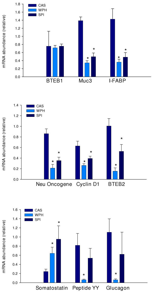
We performed quantitative real-time RT-PCR on selected genes to confirm the microarray results. Based upon known associations with cell proliferation or differentiation, 14 genes were chosen for further study. Included in this group was BTEB2; this gene was not present on the microarrays but was included in RT-PCR analysis due to its significant expression in intestine and involvement in cell proliferation [see discussion]. As shown in Figure 3, eight genes were confirmed to be differentially expressed: these included the gastrointestinal hormone genes PYY (12.9-fold down-regulated in WPH fed rats; P = 0.004), glucagon (17.8-fold down-regulated in WPH fed rats; P = 0.005), and somatostatin (3.92-, 2.65-fold up-regulated in SPI and WPH fed rats; P = 0.05, P = 0.025, respectively); cyclin D1 (1.6-, 2.4-fold down-regulated in SPI and WPH fed rats; P = 0.033, P = 0.001, respectively); BTEB2 (1.9-, 6.7-fold down-regulated in SPI and WPH fed rats; P = 0.024, P < 0.001, respectively); c-neu proto-oncogene (2.5-, 4.1-fold down-regulated in SPI and WPH fed rats; P < 0.001, P < 0.001, respectively); the colonocyte differentiation marker I-FABP (2.9-, 4.0-fold down-regulated in SPI and WPH fed rats; P = 0.023, P = 0.01, respectively); and the mucin, MUC3 (2.78-, 4.05-fold down-regulated in SPI and WPH fed rats; P < 0.001, P < 0.001, respectively). Differential expression of five other genes was not confirmed statistically, due to individual animal variation in the transcript levels; however, the mean-fold changes for mRNA abundance were greater than two and in agreement with the corresponding microarray results for these genes. Only one of the selected genes – retinol binding protein (RBP), failed to exhibit greater than a 2-fold change (in the predicted direction) at the mRNA level by real-time RT-PCR.
Figure 3.
Quantitative real-time RT-PCR verification of microarray results. RNA used for real-time RT-PCRs was from the same animals (n = 7 per diet group) whose RNAs comprised the pools for microarray analysis. Values are mean ± SEM and were analyzed by one-way ANOVA, *P < 0.05, SPI or WPH vs. CAS.
Serum somatostatin (Sst)
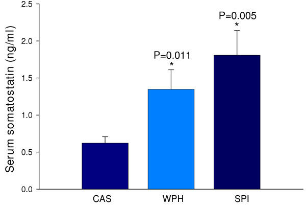
As shown in Fig. 4, circulating Sst concentration was significantly higher in rats fed WPH and SPI. Colonic Sst protein content in colon homogenates was below the limit of detection of the assay used (data not shown).
Figure 4.
Diet effects on serum Sst concentration. Values are mean ± SEM. One-way ANOVA. *P < 0.05, SPI or WPH vs. CAS.
Discussion
The type of dietary protein(s) can markedly affect the onset and/or progression of CRC [31]. Epidemiological and animal studies have found that dietary soy and whey proteins decrease the incidence of certain tumors, including those of the colon and rectum [6,7,32-35]. Using the AOM-treated male Sprague Dawley rat model, we previously found that lifetime feeding of SPI led to a ~ 76% lower incidence of AOM-induced colon tumors compared to rats lifetime-fed CAS [8]. Additionally, in the same studies, a ~ 46% lower incidence of colon tumors was found in WPH-fed compared to CAS-fed rats [9]. The molecular mechanism(s) by which these dietary proteins reduce the incidence of chemically-induced colon tumors is unclear, although several mechanisms and putative bio-active factors have been proposed [11-24]. The present study has now identified genes that are differentially expressed as a function of these diets and which serve to highlight potential pathways for dietary protection from carcinogenesis.
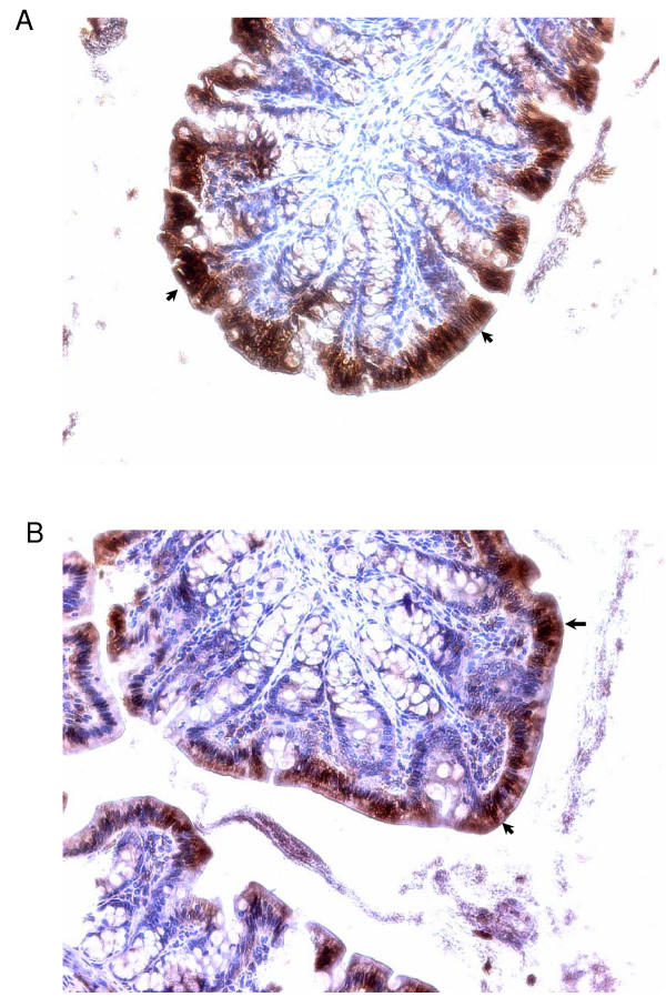
The ability to simultaneously analyze a large number of different mRNAs makes microarrays very appealing for identifying genes and gene families whose expression is altered by diet [36,37]. We focused on the 'normal' colon tissue since we are interested in genes that are differentially regulated by diet and which act in anti-oncogenic fashion in pre-cancerous tissues. We limited our analysis to the proximal colon since several studies have suggested that the molecular etiology of proximal and distal colon tumors differs [25,26] and proximal colon tumors have become more prevalent with Westernization of the diet and aging of the population [27]. We chose to include colonic smooth muscle with the mucosa since: a) the former tissue layer interacts with the latter to influence its growth and function, and b) we could monitor all colonic genes affected by diet. However, one potential caveat to this strategy is the 'dilution' effect that may have been imposed on the more rare mucosal transcripts. Another caveat is that no information is obtained regarding where the differentially expressed transcripts occur. In this regard, however, we have confirmed by immuno-histochemistry that I-FABP is expressed predominantly in the inter-cryptal surface epithelium of colons from AOM-treated rats (Fig. 5). Our study used a sample size of three (per diet group) which balanced the costs for the experimental reagents with the minimum number required for statistical analysis. The quantitative PCR analyses provided confirmation that the filtering strategies used yielded bona-fide differentially expressed transcripts.
Figure 5.
Immuno-histochemistry for I-FABP in colons from AOM-treated rats. Panels A and B are sections from CAS and WPH-fed animals, respectively. Arrows point to the strong areas of staining for I-FABP in the inter-cryptal surface epithelium (overall intensity of staining is greater for CAS than for WPH).
Only two transcripts were induced by both SPI and WPH; whereas 37 transcripts were repressed by both SPI and WPH. This suggests that the cancer-protective actions of the two diets are generally associated with repression of colonic genes that facilitate tumorigenesis. An alternative explanation is that CAS induces genes that facilitate colon cancer development when compared to SPI and WPH. It is also likely that SPI and WPH diets act in unique ways to inhibit tumorigenesis. Irregardless, our results indicate that the nature of the dietary protein can profoundly affect colon gene expression profiles. Thus, gene expression profiling studies of colons should account for potential confounding effects of diet.
Dietary factors in SPI or WPH inhibit cell proliferation and induce apoptosis among other biological actions [11,13]. In the present study, we identified cyclin D1 gene and the neu proto-oncogene as being repressed in proximal colon by SPI and WPH. Cyclin D1 is a key regulator of cell cycle progression [38], and a target of β-catenin, a protein whose abnormal accumulation in the nucleus is strongly linked to the development of multiple tumor types, including those of the colon [39]. Aberrantly increased expression of cyclin D1 in colon epithelial cells contributes to their abnormal proliferation and tumorigenicity [40,41]. Similarly, the oncogenic and cellular growth-promoting activities of the HER-2/neu proto-oncogene are well known [42]. HER-2/neu, a tyrosine kinase receptor for neu-differentiation factor, is expressed in normal colonic epithelium and is up-regulated in adenomatous polyps of the colon [43]. The down regulation of cyclin D1 and c-neu mRNA abundance by SPI and WPH may at least partly explain their anti-tumorigenic properties. Similarly, Krüppel-like transcription factors have been linked to cell growth and tumorigenesis. BTEB2 (also known as Krüppel-like factor 5, KLF5, or intestinal KLF) was reported to enhance intestinal epithelia cell colony formation, cyclin D1 transcription, and cell proliferation [44]. Consistent with our results for cyclin D1, colonic BTEB2 mRNA expression was down-regulated by SPI and WPH. Aquaporin 3, a water channel highly expressed in colonic epithelium, was down-regulated by SPI and WPH. Aquaporins are thought to be induced early in colon cancer and to facilitate oncogenesis [45], therefore, dietary repression of such genes may additionally contribute to anti-tumorigenesis. The results for I-FABP and MUC3 indicated 3–4 fold decreases in transcript abundance in proximal colons of rats on SPI or WPH diets. These particular results are not easily reconciled with decreased tumorigenesis in SPI and WPH groups, since both genes are highly expressed in the normal differentiated colonic epithelium and are likely to be under-expressed in adenomas and adenocarcinomas [46]. Perhaps, these represent diet-modulated genes that are not direct participants in anti-tumorigenesis.
Gastrointestinal hormones regulate a myriad of intestinal functions including motility, absorption, digestion, cell proliferation and death, and immune response [47]. The microarray and real-time RT-PCR assays identified inductive effects of SPI and WPH on somatostatin mRNA and protein abundance. These results implicate this gene product in autocrine and paracrine mechanisms underlying colon cancer protection by SPI and WPH since somatostatin is a well-known anti-proliferative agent for colon tumor cells [48,49]. This hormone is also a negative regulator of angiogenesis [50]; this is predicted to counter tumorigenesis. It is possible that the small decrements in rat growth rates observed with lifetime SPI or WPH diets [8,9] are a consequence of this increased circulating somatostatin level. We also found decreased abundance of mRNAs encoding peptide YY (PYY) and glucagon in colons of WPH-fed rats. PYY gene expression in human colon tumors is much reduced relative to the adjacent normal tissue [51]; however, chemically-induced colon tumors in rats generally exhibit higher overall expression of PYY due to increased prevalence of PYY-positive cells, compared to normal mucosa [52,53]. PYY stimulates proliferation of intestinal epithelium [54]; therefore, an inhibition of PYY expression by dietary WPH may contribute to colon cancer-protective actions. A similar scenario might apply to colon glucagon gene expression, as this growth stimulatory peptide for colon cancer cells [55] was inhibited by WPH at the level of colon mRNA abundance.
Our data highlighted other aspects of diet and colon gene expression that warrant further study. For example, the B7 antigen (also known as CD52) mRNA was strongly down-regulated by SPI or WPH. The corresponding protein is normally expressed at high levels on cell membranes of T and B lymphocytes and monocytes; infusion with anti-CD52 antibody leads to systemic depletion of T cells [56]. The lower abundance of this transcript in non-tumor colon tissue of rats on SPI or WPH diets may reflect fewer numbers of immune cells in this tissue, as compared to CAS-fed animals. One possible interpretation of this data is that the 'normal' tissue of the CAS group has manifested an immune response, perhaps in response to increased tumorigenicity relative to SPI or WPH groups. Such an interpretation raises the prospect of a functional immuno-editing mechanism [57] occurring in this model of colon cancer and an indirect effect of diet on lymphocyte populations (via presence of tumors or tumor precursors) in the colon. An alternative mechanism is that dietary protein can directly affect the populations of lymphocytes resident in the colon, which in turn, may affect tumorigenesis. A related observation was the enhanced abundance of CD3 gamma chain transcripts in colons of SPI and WPH animals. The protein encoded by this transcript helps mediate T cell antigen receptor engagement and signaling [58]; its decreased abundance in colonic T cells of CAS-fed animals may indicate a specific immune defect [59] occurring in the CAS-fed animals after exposure to carcinogen and thereby contributing to enhanced tumorigenesis in this group.
Several microarray studies of human paired normal colon vs. colon tumors have been published [60-64]. Comparison of the present results for normal colon tissue of AOM-treated rats on different diets to the published studies for human CRC identified only a small number of common differentially expressed genes and/or gene families in common (data not shown). This small number is probably due to the fact that our study did not examine colon tumors; rather we focused on 'normal' colon tissue. In this regard, it will be interesting to examine the expression profiles of colons from animals not treated with AOM so as to more specifically correlate transcripts with diet and cancer phenotype. This study has illuminated a number of genes and gene families that may act as dietary protein-dependent modulators of oncogenesis in the rat colon. Additional studies that specifically address the functional involvement of these genes in cancer-prevention via dietary means are required to confirm the postulated roles.
Conclusions
We have identified genes in rat colon that are differentially expressed, as a consequence of altered dietary protein, during AOM-induced oncogenesis. These are candidates for genes that sub-serve the anti-cancer effects of dietary SPI and WPH in this tissue.
Methods
Rats, diets and carcinogen treatment
The animals whose colons were used in the present study have been previously described [8,9]. Time-mated [gestation day (GD) 4] Sprague-Dawley rats were purchased from Harlan Industries (Indianapolis, IN), housed individually and allowed ad libitum access to water and pelleted food. Rats were randomly assigned to one of three semi-purified isocaloric diets made according to the AIN-93G diet formula [65] and which differed only by protein source: a) CAS (New Zealand Milk Products, Santa Rosa, CA), b) WPH (New Zealand Milk Products, Santa Rosa, CA) or c) SPI (Dupont Protein Technologies, St. Louis, MO). Offspring were weaned to the same diet as their mothers and were fed the same diets throughout the study. At 90 days of age, male offspring received s.c. injections of 15 mg/kg AOM (Ash Stevens, Detroit, MI) in saline once a week for 2 weeks. Forty weeks later, rats were euthanized, and the colon (cecum to anus) was divided into two equal segments (proximal and distal), opened longitudinally, and examined for tumors. We found that both WPH and SPI significantly decreased the colon tumor incidence [data published in [8,9]]. A representative non-tumor segment of each proximal colon (PC) was frozen in liquid nitrogen and stored at -80°C for later use. Animal care and handling were in accordance with the Institutional Animal Care & Use Committee guidelines of the University of Arkansas for Medical Sciences.
RNA processing
Total RNA was isolated from rat proximal colons (n = 7 for each of CAS, SPI and WPH diets) using TRIzol reagent (Invitrogen, Carlsbad, CA), and further purified with the RNeasy Mini Kit (QIAGEN, Valencia, CA). To remove contaminating DNA, on-column DNA digestion with RNase-Free DNase (QIAGEN) was performed. Integrity of isolated RNAs was confirmed using the RNA 6000 Nano LabChip kit with the Agilent 2100 Bioanalyzer System (Agilent Biotechnologies, Palo Alto, CA). To reduce errors due to biological variability, RNA samples were pooled as proposed by Bakay et al [66]. Pooled RNA (equal amounts of RNA from each of 7 animals; 8 ug total) was used for cDNA synthesis using a T7-(deoxythymidine)24 primer and Superscript II (Life Technologies, Inc., Gaithersburg, MD). The resulting cDNA was used with the ENZO BioArray High Yield RNA Transcript labeling kit (ENZO, Farmingdale, NY) to synthesize biotin-labeled cRNA. The cRNA was purified on RNeasy spin columns (QIAGEN) and subjected to chemical fragmentation (size range of 35 to 200 bp). Three replicate cRNA targets were made in parallel starting from each RNA pool.
Microarray procedures
Ten ug of cRNA was hybridized for 16 hours to an Affymetrix (Santa Clara, CA) rat U34A GeneChip (3 chips used per diet group), followed by incubations with streptavidin-conjugated phycoerythrin, and then with polyclonal anti-streptavidin antibody coupled to phycoerythrin. Following washing, GeneChips were scanned using an Agilent GeneArray laser scanner. Images were analyzed using Affymetrix MAS 5.0 software. Bacterial sequence-derived probes on the arrays served as external controls for hybridization, whereas the housekeeping genes β-actin and GAPDH served as endogenous controls and for monitoring the quality of the RNA target. To compare array data between GeneChips, we scaled the average of the fluorescent intensities of all probes on each array to a constant target intensity of 500.
Bioinformatics
To validate the microarray procedure for our samples, unsupervised nearest-neighbor hierarchical clustering (Spotfire, Somerville, MA) was performed on gene expression data. The inter-chip variability test also was performed as specified in the Affymetrix data analysis manual [28]. To identify colon genes differentially expressed with SPI or WPH (control: CAS diet), multiple criteria were applied; final results are reported only for transcripts that passed all three analytical steps described below. Firstly, the t-test feature of DMT (Affymetrix) was used to identify genes whose expression was regulated (induced/repressed with P < 0.05) by SPI or WPH, and signal fold changes (FC) for these genes were calculated. Secondly, microarray data were analyzed using 'Significance of Analysis of Microarrays' (SAM, Stanford) to identify significant changes in gene expression among diet groups [67], using a false discovery rate (FDR) cutoff of 0.5%. Lastly, a pair-wise comparison survival (3 × 3) method was used to identify differentially expressed transcripts [68]. In brief, the three replicate expression profiles obtained for SPI colons were iteratively compared with the three CAS profiles (latter as baseline) in MAS 5.0 (Affymetrix), generating nine comparisons in total. Transcripts with a log ratio greater than or equal to 1 (≥2 fold change), which increased (I) in nine of nine comparisons, and which were expressed above background (i.e., called as Present) in all three SPI GeneChips, were considered to be up-regulated by SPI. Transcripts with a log ratio less than or equal to -1, were decreased (D) in nine of nine comparisons, and expressed above background (Present) in all three CAS chips were considered to be down-regulated by SPI. WPH-regulated genes were similarly identified. Genes that were independently identified by all three approaches comprised the final reported lists of differentially expressed genes (Tables 2, 3, 4, 5, 6).
Validation of gene expression by quantitative real-time RT-PCR
One μg of total RNA from each of the 21 individual proximal colons (which comprised the original pools for the microarray experiment) was reverse-transcribed using random hexamers and MultiScribe Reverse Transcriptase in a two-step RT-PCR reaction (Applied Biosystems, Foster City, CA). Primers (Table 7) were designed using 'Primer Express' (Applied Biosystems) and were selected to yield a single amplicon; this was verified by dissociation curves and/or analysis in agarose gels. SYBR Green real-time PCR was performed with an ABI Prism 7000 Sequence Detector. Thermal cycling conditions included pre-incubation at 50°C for 2 min, 95°C for 10 min followed by 40 PCR cycles at 95°C for 15 sec and 60°C for 1 min. The relative transcript levels for each gene were calculated using the relative standard curve method (User Bulletin #2, Applied Biosystems) and normalized to the house-keeping gene β-actin. Data are reported as mean ± SEM of n = 7 animals per dietary group. Significant differences between diet groups were determined by one-way ANOVA (P < 0.05).
Table 7.
Primer sequences for real-time RT-PCR
| Gene | Forward primer | Reverse primer | Accession no. |
| Beta-actin | 5'-GACGGTCAGGTCATCACTATCG-3' | 5'-ACGGATGTCAACGTCACACTTC-3' | NM_031144 |
| I-FABP | 5'-AGGAAGCTTGGAGCTCATGACA-3' | 5'-TCCTTCCTGTGTGATCGTCAGTT-3' | K01180 |
| Neu Oncogene | 5'-GTGGTCGTTGGAATCCTAATCAA-3' | 5'-CCTTCCTTAGCTCCGTCTCTTTTA-3' | X03362 |
| PYY | 5'-AGGAGCTGAGCCGCTACTATGC-3' | 5'-TTCTCGCTGTCGTCTGTGAAGA-3' | M17523 |
| Glucagon | 5'-TGGTGAAAGGCCGAGGAAG-3' | 5'-TGGTGGCAAGGTTATCGAGAA-3' | K02813 |
| Somatostatin | 5'-GGAAACAGGAACTGGCCAAGT-3' | 5'-TGCAGCTCCAGCCTCATCTC-3' | K02248 |
| PAP III | 5'-AAGAGGCCATCAGGACACCTT-3' | 5'-CACTCCCATCCACCTCTGTTG-3' | L20869 |
| CYP4F1 | 5'-CCAAGTGGAAACGGTTGATTTC-3' | 5'-TCCTGGCAGTTGCTGTCAAAG-3' | M94548 |
| GST | 5'-ACTTCCCCAATCTGCCCTACTTA-3' | 5'-CGAATCCGCTCCTCCTCTGT-3' | X04229 |
| Cyclin D1 | 5'-TCAAGTGTGACCCGGACTGC-3' | 5'-ACTTCCCCTTCCTCCTCGGT-3' | D14014 |
| Beta defensin-1 | 5'-TCTTGGACGCAGAACAGATCAATA-3' | 5'-TCCTGCAACAGTTGGGCTATC-3' | AF093536 |
| H+, K+-ATPase | 5'-ATTCCGCATCCCTAGACAACG-3' | 5'-TCTTACTAAAGCTGGCCATGATGTT-3' | M90398 |
| Prostaglandin D synthetase | 5'-CAAGCTGGTTCCGGGAGAAG-3' | 5'-TTGGTCTCACACTGGTTTTTCCTTA-3' | J04488 |
| RBP | 5'-TCGTTTCTCTGGGCTCTGGTAT- 3' | 5'-TTCCCAGTTGCTCAGAAGACG-3' | M10934 |
| Muc3 | 5'-AAGGTGTGAGGAAGTGATGGAGA-3' | 5'-GCAGAGACCGTCGGCTTTATC-3' | U76551 |
| BTEB1 | 5'-ACACTGGTCACCATCGCCAA-3' | 5'-GGACTCGACCCAGATTCGGT-3' | NM_057211 |
| BTEB2 | 5'-CTACTTTCCCCCATCACCACC-3' | 5'-GAATCGCCAGTTTCGAAGCA-3' | AB096709 |
Serum Sst
Rat serum Sst content (15 animals from each diet) was determined using the somatostatin-28 EIA kit purchased from Phoenix Pharmaceuticals Corporation (Belmont, California).
Authors' contributions
RX performed the microarray and real-time PCR experiments, conducted the data analysis, and participated in drafting the manuscript. TMB designed and oversaw the animal component of the study. FAS designed the analytical and overall approaches to the study, supervised the project, and drafted the manuscript. All authors read and approved the final manuscript.
Acknowledgments
Acknowledgements
We thank Dr. Rosalia C.M. Simmen and Dr. Rick Helm for insightful comments on the manuscript and Amanda L. Linz for performing the I-FABP immuno-histochemistry. Supported by USDA CRIS 6251-5100-002-06S.
Contributor Information
Rijin Xiao, Email: XiaoRijin@uams.edu.
Thomas M Badger, Email: BadgerThomasM@uams.edu.
Frank A Simmen, Email: SimmenFrankA@uams.edu.
References
- Cancer statistics 2004 – a presentation from the American Cancer Society http://www. cancer.org/downloads/PRO/Cancer%20Statistics%202004.ppt
- A snapshot of colorectal cancer http://prg.nci.nih.gov/snapshots/Colorectal-Snapshot.pdf
- Sandler RS. Epidemiology and risk factors for colorectal cancer. Gastroenterol Clin North Am. 1996;25:717–735. doi: 10.1016/s0889-8553(05)70271-5. [DOI] [PubMed] [Google Scholar]
- Potter JD. Nutrition and colorectal cancer. Cancer Causes Control. 1996;7:127–146. doi: 10.1007/BF00115644. [DOI] [PubMed] [Google Scholar]
- Levi F, Pasche C, La Vecchia C, Lucchini F, Franceschi S. Food groups and colorectal cancer risk. Br J Cancer. 1999;79:1283–1287. doi: 10.1038/sj.bjc.6690206. [DOI] [PMC free article] [PubMed] [Google Scholar]
- Corpet DE, Tache S. Most effective colon cancer chemopreventive agents in rats: a systematic review of aberrant crypt foci and tumor data, ranked by potency. Nutr Cancer. 2002;43:1–21. doi: 10.1207/S15327914NC431_1. [DOI] [PMC free article] [PubMed] [Google Scholar]
- Kelloff GJ, Crowell JA, Steele VE, Lubet RA, Boone CW, Malone WA, Hawk ET, Lieberman R, Lawrence JA, Kopelovich L, Ali I, Viner JL, Sigman CC. Progress in cancer chemoprevention. Ann N Y Acad Sci. 1999;889:1–13. doi: 10.1111/j.1749-6632.1999.tb08718.x. [DOI] [PubMed] [Google Scholar]
- Hakkak R, Korourian S, Ronis MJ, Johnston JM, Badger TM. Soy protein isolate consumption protects against azoxymethane-induced colon tumors in male rats. Cancer Lett. 2001;166:27–32. doi: 10.1016/S0304-3835(01)00441-4. [DOI] [PubMed] [Google Scholar]
- Hakkak R, Korourian S, Ronis MJ, Johnston JM, Badger TM. Dietary whey protein protects against azoxymethane-induced colon tumors in male rats. Cancer Epidemiol Biomarkers Prev. 2001;10:555–558. [PubMed] [Google Scholar]
- Belobrajdic DP, McIntosh GH, Owens JA. Whey proteins protect more than red meat against azoxymethane induced ACF in Wistar rats. Cancer Lett. 2003;198:43–51. doi: 10.1016/S0304-3835(03)00307-0. [DOI] [PubMed] [Google Scholar]
- Sternhagen LG, Allen JC. Growth rates of a human colon adenocarcinoma cell line are regulated by the milk protein alpha-lactalbumin. Adv Exp Med Biol. 2001;501:115–120. doi: 10.1007/978-1-4615-1371-1_14. [DOI] [PubMed] [Google Scholar]
- Guo JY, Li X, Browning JD, Jr, Rottinghaus GE, Lubahn DB, Constantinou A, Bennink M, MacDonald RS. Dietary soy isoflavones and estrone protect ovariectomized ERα KO and wild-type mice from carcinogen-induced colon cancer. J Nutr. 2004;134:179–182. doi: 10.1093/jn/134.1.179. [DOI] [PubMed] [Google Scholar]
- Yanagihara K, Ito A, Toge T, Numoto M. Antiproliferative effects of isoflavones on human cancer cell lines established from the gastrointestinal tract. Cancer Res. 1993;53:5815–5821. [PubMed] [Google Scholar]
- Booth C, Hargreaves DF, Hadfield JA, McGown AT, Potten CS. Isoflavones inhibit intestinal epithelial cell proliferation and induce apoptosis in vitro. Br J Cancer. 1999;80:1550–1557. doi: 10.1038/sj.bjc.6690559. [DOI] [PMC free article] [PubMed] [Google Scholar]
- Wenzel U, Kuntz S, Brendel MD, Daniel H. Dietary flavone is a potent apoptosis inducer in human colon carcinoma cells. Cancer Res. 2000;60:3823–3831. [PubMed] [Google Scholar]
- Booth C, Hargreaves DF, O'Shea JA, Potten CS. In vivo administration of genistein has no effect on small intestinal epithelial proliferation and apoptosis, but a modest effect on clonogen survival. Cancer Lett. 1999;144:169–175. doi: 10.1016/S0304-3835(99)00220-7. [DOI] [PubMed] [Google Scholar]
- Symolon H, Schmelz EM, Dillehay DL, Merrill AL., Jr Dietary soy sphingolipids suppress tumorigenesis and gene expression in 1,2-dimethylhydrazine-treated CF1 mice and ApcMin/+ mice. J Nutr. 2004;134:1157–1161. doi: 10.1093/jn/134.5.1157. [DOI] [PubMed] [Google Scholar]
- Rowlands JC, He L, Hakkak R, Ronis MJ, Badger TM. Soy and whey proteins downregulate DMBA-induced liver and mammary gland CYP1 expression in female rats. J Nutr. 2001;131:3281–3287. doi: 10.1093/jn/131.12.3281. [DOI] [PubMed] [Google Scholar]
- Appelt LC, Reicks MM. Soy feeding induces phase II enzymes in rat tissues. Nutr Cancer. 1997;28:270–275. doi: 10.1080/01635589709514587. [DOI] [PubMed] [Google Scholar]
- Appelt LC, Reicks MM. Soy induces phase II enzymes but does not inhibit dimethylbenz[a]anthracene-induced carcinogenesis in female rats. J Nutr. 1999;129:1820–1826. doi: 10.1093/jn/129.10.1820. [DOI] [PubMed] [Google Scholar]
- McIntosh GH, Regester GO, Le Leu RK, Royle PJ, Smithers GW. Dairy proteins protect against dimethylhydrazine-induced intestinal cancers in rats. J Nutr. 1995;125:809–816. doi: 10.1093/jn/125.4.809. [DOI] [PubMed] [Google Scholar]
- Bounous G, Batist G, Gold P. Whey proteins in cancer prevention. Cancer Lett. 1991;57:91–94. doi: 10.1016/0304-3835(91)90200-2. [DOI] [PubMed] [Google Scholar]
- Bounous G. Whey protein concentrate (WPC) and glutathione modulation in cancer treatment. Anticancer Res. 2000;20:4785–4792. [PubMed] [Google Scholar]
- Wargovich MJ. Colon cancer chemoprevention with ginseng and other botanicals. J Korean Med Sci. 2001;16:S81–86. doi: 10.3346/jkms.2001.16.S.S81. [DOI] [PMC free article] [PubMed] [Google Scholar]
- Distler P, Holt PR. Are right- and left-sided colon neoplasms distinct tumors? Dig Dis. 1997;15:302–311. doi: 10.1159/000171605. [DOI] [PubMed] [Google Scholar]
- Hong MY, Chapkin RS, Morris JS, Wang N, Carroll RJ, Turner ND, Chang WC, Davidson LA, Lupton JR. Anatomical site-specific response to DNA damage is related to later tumor development in the rat azoxymethane colon carcinogenesis model. Carcinogenesis. 2001;22:1831–1835. doi: 10.1093/carcin/22.11.1831. [DOI] [PubMed] [Google Scholar]
- Cady B, Stone MD, Wayne J. Continuing trends in the prevalence of right-sided lesions among colorectal carcinomas. Arch Surg. 1993;128:505–509. doi: 10.1001/archsurg.1993.01420170035004. [DOI] [PubMed] [Google Scholar]
- Affymetrix, GeneChip expression Analysis – Data analysis fundamentals (Manual) 2002.
- Sohn OS, Fiala ES, Requeijo SP, Weisburger JH, Gonzalez FJ. Differential effects of CYP2E1 status on the metabolic activation of the colon carcinogens azoxymethane and methylazoxymethanol. Cancer Res. 2001;61:8435–8440. [PubMed] [Google Scholar]
- Fruchart JC, De Geteire C, Delfly B, Castro GR. Apolipoprotein A-I-containing particles and reverse cholesterol transport: evidence for connection between cholesterol efflux and atherosclerosis risk. Atherosclerosis. 1994;110:S35–S39. doi: 10.1016/0021-9150(94)05374-r. [DOI] [PubMed] [Google Scholar]
- Adlercreutz CH, Goldin SL, Gorbach KA, Hockerstedt S, Watanabe EK, Hamalainen MH, Makkanen TH, Wahala KT, Adlercreutz T. Soybean phytoestrogen intake and cancer risk. J Nutr. 1995;125:757S–770S. doi: 10.1093/jn/125.3_Suppl.757S. [DOI] [PubMed] [Google Scholar]
- Tsuda H, Sekine K, Takasuka N, Toriyama-Baba H, Iigo M. Prevention of colon carcinogenesis and carcinoma metastasis by orally administered bovine lactoferrin in animals. Biofactors. 2000;12:83–88. doi: 10.1002/biof.5520120113. [DOI] [PubMed] [Google Scholar]
- Papenburg R, Bounous G, Fleiszer D, Gold P. Dietary milk proteins inhibit the development of dimethylhydrazine-induced malignancy. Tumour Biol. 1990;11:129–136. doi: 10.1159/000217647. [DOI] [PubMed] [Google Scholar]
- Toyomura K, Kono S. Soybeans, soy foods, isoflavones and risk of colorectal cancer: a review of experimental and epidemiological data. Asian Pac J Cancer Prev. 2002;3:125–132. [PubMed] [Google Scholar]
- Spector D, Anthony M, Alexander D, Arab L. Soy consumption and colorectal cancer. Nutr Cancer. 2003;47:1–12. doi: 10.1207/s15327914nc4701_1. [DOI] [PubMed] [Google Scholar]
- Deaciuc IV, Peng X, D'Souza NB, Shedlofsky SI, Burikhanov R, Voskresensky IV, de Villiers WJ. Microarray gene analysis of the liver in a rat model of chronic, voluntary alcohol intake. Alcohol. 2004;32:113–127. doi: 10.1016/j.alcohol.2003.12.001. [DOI] [PubMed] [Google Scholar]
- Kato N, Shibutani M, Takagi H, Uneyama C, Lee KY, Takigami S, Mashima K, Hirose M. Gene expression profile in the livers of rats orally administered ethinylestradiol for 28 days using a microarray technique. Toxicology. 2004;200:179–192. doi: 10.1016/j.tox.2004.03.008. [DOI] [PubMed] [Google Scholar]
- Charles JS. Cancer Cell Cycles. Science. 1996;274:1672–1677. doi: 10.1126/science.274.5293.1672. [DOI] [PubMed] [Google Scholar]
- Tetsu O, McCormick F. β-Catenin regulates expression of cyclin D1 in colon carcinoma cells. Nature. 1999;398:422–426. doi: 10.1038/18884. [DOI] [PubMed] [Google Scholar]
- Arber N, Doki Y, Han EK-H, Sgambato A, Zhou P, Kim N-H, Delohery T, Klein MG, Holt PR, Weinstein IB. Antisense to cyclin D1 inhibits the growth and tumorigenicity of human colon cancer cells. Cancer Res. 1997;57:1569–1574. [PubMed] [Google Scholar]
- Al Moustafa AE, Foulkes WD, Wong A, Jallal H, Batist G, Yu Q, Herlyn M, Sicinski P, Alaoui-Jamali MA. Cyclin D1 is essential for neoplastic transformation induced by both E6/E7 and E6/E7/ErbB-2 cooperation in normal cells. Oncogene. 2004;23:5252–5256. doi: 10.1038/sj.onc.1207679. [DOI] [PubMed] [Google Scholar]
- De Potter CR. The neu oncogene: more than a prognostic indicator? Hum Pathol. 1994;25:1264–1268. doi: 10.1016/0046-8177(94)90083-3. [DOI] [PubMed] [Google Scholar]
- Cohen JA, Weiner DB, More KF, Kokai Y, Williams WV, Maguire HC, Jr, LiVolsi VA, Greene MI. Expression pattern of the neu (NGL) gene-encoded growth factor receptor protein (p185neu) in normal and transformed epithelial tissues of the digestive tract. Oncogene. 1989;4:81–88. [PubMed] [Google Scholar]
- Bateman NW, Tan D, Pestell PG, Black JD, Black AR. Intestinal tumor progression is associated with altered function of KLF5. J Biol Chem. 2004;279:12093–12101. doi: 10.1074/jbc.M311532200. [DOI] [PubMed] [Google Scholar]
- Moon C, Soria JC, Jang SJ, Lee J, Obaidul Hoque M, Sibony M, Trink B, Chang YS, Sidransky D, Mao L. Involvement of aquaporins in colorectal carcinogenesis. Oncogene. 2003;22:6699–6703. doi: 10.1038/sj.onc.1206762. [DOI] [PubMed] [Google Scholar]
- Chang SK, Dohrman AF, Basbaum CB, Ho SB, Tsuda T, Toribara NW, Gum JR, Kim YS. Localization of mucin (MUC2 and MUC3) messenger RNA and peptide expression in human normal intestine and colon cancer. Gastroenterology. 1994;107:28–36. doi: 10.1016/0016-5085(94)90057-4. [DOI] [PubMed] [Google Scholar]
- Thomas RP, Hellmich MR, Townsend CM, Jr, Evers BM. Role of gastrointestinal hormones in the proliferation of normal and neoplastic tissues. Endocr Rev. 2003;24:571–599. doi: 10.1210/er.2002-0028. [DOI] [PubMed] [Google Scholar]
- Bousquet C, Puente E, Buscail L, Vaysse N, Susini C. Antiproliferative effect of somatostatin and analogs. Chemotherapy. 2001;47:30–39. doi: 10.1159/000049159. [DOI] [PubMed] [Google Scholar]
- Tejeda M, Gaal D, Barna K, Csuka O, Keri G. The antitumor activity of the somatostatin structural derivative (TT-232) on different human tumor xenografts. Anticancer Res. 2003;23:4061–4066. [PubMed] [Google Scholar]
- Florio T, Morini M, Villa V, Arena S, Corsaro A, Thellung S, Culler MD, Pfeffer U, Noonan DM, Schettini G, Albini A. Somatostatin inhibits tumor angiogenesis and growth via somatostatin receptor-3-mediated regulation of endothelial nitric oxide synthase and mitogen-activated protein kinase activities. Endocrinology. 2003;144:1574–1584. doi: 10.1210/en.2002-220949. [DOI] [PubMed] [Google Scholar]
- Adrian TE, Ballantyne GH, Zucker KA, Zdon MJ, Tierney R, Modlin IM. Lack of peptide YY immunoreactivity in adenomatous colonic polyps: evidence in favor of an adenoma-carcinoma sequence. J Surg Res. 1988;44:561–565. doi: 10.1016/0022-4804(88)90162-X. [DOI] [PubMed] [Google Scholar]
- Johnston CF, O'Neill AB, O'Hare MM, Buchanan KD. Neuroendocrine cells within colorectal tumours induced by dimethylhydrazine. An immunocytochemical study. Cell Tissue Res. 1986;246:205–210. doi: 10.1007/BF00219019. [DOI] [PubMed] [Google Scholar]
- Sitohy B, El-Salhy M. Colonic endocrine cells in rats with chemically induced colon carcinoma. Histol Histopathol. 2001;16:833–838. doi: 10.14670/HH-16.833. [DOI] [PubMed] [Google Scholar]
- Mannon PJ. Peptide YY as a growth factor for intestinal epithelium. Peptides. 2002;23:383–388. doi: 10.1016/S0196-9781(01)00615-5. [DOI] [PubMed] [Google Scholar]
- Moyer MP, Aust JB, Dixon PS, Levine BA, Sirinek KR. Glucagon enhances growth of cultured human colorectal cancer cells in vitro. Am J Surg. 1985;150:676–679. doi: 10.1016/0002-9610(85)90406-4. [DOI] [PubMed] [Google Scholar]
- Brett SJ, Baxter G, Cooper H, Rowan W, Regan T, Tite J, Rapson N. Emergence of CD52-, glycosylphosphatidylinositol-anchor-deficient lymphocytes in rheumatoid arthritis patients following Campath-1H treatment. Int Immunol. 1996;8:325–334. doi: 10.1093/intimm/8.3.325. [DOI] [PubMed] [Google Scholar]
- Dunn GP, Old LJ, Schreiber RD. The three Es of cancer immunoediting. Annu Rev Immunol. 2004;22:329–360. doi: 10.1146/annurev.immunol.22.012703.104803. [DOI] [PubMed] [Google Scholar]
- Pitcher LA, van Oers NS. T-cell receptor signal transmission: who gives an ITAM? Trends Immunol. 2003;24:554–560. doi: 10.1016/j.it.2003.08.003. [DOI] [PubMed] [Google Scholar]
- Mizoguchi H, O'Shea JJ, Longo DL, Loeffler CM, McVicar DW, Ochoa AC. Alterations in signal transduction molecules in T lymphocytes from tumor-bearing mice. Science. 1992;258:1795–1798. doi: 10.1126/science.1465616. [DOI] [PubMed] [Google Scholar]
- Kitahara O, Furukawa Y, Tanaka T, Kihara C, Ono K, Yanagawa R, Nita ME, Takagi T, Nakamura Y, Tsunoda T. Alterations of gene expression during colorectal carcinogenesis revealed by cDNA microarrays after laser-capture microdissection of tumor tissues and normal epithelia. Cancer Res. 2001;61:3544–3549. [PubMed] [Google Scholar]
- Notterman DA, Alon U, Sierk AJ, Levine AJ. Transcriptional gene expression profiles of colorectal adenoma, adenocarcinoma, and normal tissue examined by oligonucleotide arrays. Cancer Res. 2001;61:3124–3130. [PubMed] [Google Scholar]
- Agrawal D, Chen T, Irby R, Quackenbush J, Chambers AF, Szabo M, Cantor A, Coppola D, Yeatman TJ. Osteopontin identified as lead marker of colon cancer progression, using pooled sample expression profiling. J Natl Cancer Inst. 2002;94:513–521. doi: 10.1093/jnci/94.7.513. [DOI] [PubMed] [Google Scholar]
- Birkenkamp-Demtroder K, Christensen LL, Olesen SH, Frederiksen CM, Laiho P, Aaltonen LA, Laurberg S, Sorensen FB, Hagemann R, Orntoft TF. Gene expression in colorectal cancer. Cancer Res. 2002;62:4352–4363. [PubMed] [Google Scholar]
- Williams NS, Gaynor RB, Scoggin S, Verma U, Gokaslan T, Simmang C, Fleming J, Tavana D, Frenkel E, Becerra C. Identification and validation of genes involved in the pathogenesis of colorectal cancer using cDNA microarrays and RNA interference. Clin Cancer Res. 2003;9:931–946. [PubMed] [Google Scholar]
- Reeves PG, Nielsen FH, Fahey GC., Jr AIN-93 purified diets for laboratory rodents: final report of the American Institute of Nutrition ad hoc writing committee on the reformulation of the AIN-76A rodent diet. J Nutr. 1993;123:1939–1951. doi: 10.1093/jn/123.11.1939. [DOI] [PubMed] [Google Scholar]
- Bakay M, Chen YW, Borup R, Zhao P, Nagaraju K, Hoffman EP. Sources of variability and effect of experimental approach on expression profiling data interpretation. BMC Bioinformatics. 2002;3:4. doi: 10.1186/1471-2105-3-4. [DOI] [PMC free article] [PubMed] [Google Scholar]
- Tusher VG, Tibshirani R, Chu G. Significance analysis of microarrays applied to the ionizing radiation response. Proc Natl Acad Sci USA. 2001;98:5116–5121. doi: 10.1073/pnas.091062498. [DOI] [PMC free article] [PubMed] [Google Scholar]
- Chen Y, Zhao P, Borup R, Hoffman EP. Expression profiling in the muscular dystrophies: identification of novel aspects of molecular pathophysiology. J Cell Biol. 2000;151:1321–1336. doi: 10.1083/jcb.151.6.1321. [DOI] [PMC free article] [PubMed] [Google Scholar]


