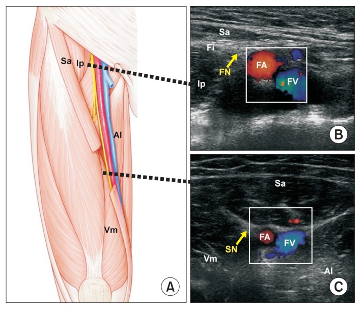
Fig. 2.
(A) Schematic drawing of the anterior aspect of right thigh. The mid portion of the sartorius muscle was cut to show the inside of the adductor canal. (B) Cross-sectional ultrasonography image at the apex of the femoral triangle. (C) Cross-sectional ultrasonography image of the adductor canal. FN: femoral nerve, FA: femoral artery, FV: femoral vein, SN: saphenous nerve, Sa: sartorius muscle, Ip: iliopsoas muscle, Fi: fascia iliaca, Vm: vatus medialis muscle, Al: adductor longus muscle.

