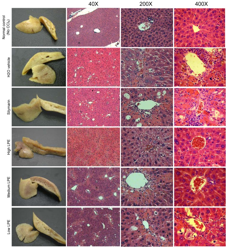
Figure 3.
The gross appearance and microscopic histopathology (haematoxylin and eosin staining; magnification, ×40, ×200, and ×400) of liver tissue from the normal animal group (control) and treatment groups of silymarin, H2O, high-dose LPE (200 mg/kg BW/day), middle-dose LPE (100 mg/kg BW/day), and low-dose LPE (20 mg/kg BW/day) in mice with CCl4-induced liver injury. Based on the gross appearance of the liver, the normal and high LPE groups had similar smooth outer capsules compared with those of other groups. Histopathological analysis demonstrated the hepatocyte edges formed pentagonal lobule architecture were retained in all treatments; however, steatosis can be seen in the silymarin, H2O, medium-LPE and low-LPE groups (magnification, ×40). CCl4-intoxication resulted in swollen hepatocytes and impaired lipid synthesis, resulting in the formation of a fatty vacuole (macrosteatosis) in the low-level LPE, silymarin and H2O vehicle-control groups (magnification, ×200). Furthermore, eosinophil infiltration (eosinophilia) along the central portal veins extending to the lobular venous tract and dark-purple/blue stained Kupffer cells infiltration along the central portal vein were evident in the in the low-LPE, silymarin and H2O groups. However, fewer focal observations were noted in the middle and high-LPE samples (magnification, ×400). LPE, litchi pericarp extract; BW, body weight; CCl4, carbon tetracholoride.

