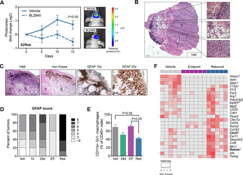
Figure 3. Resistance to CSF-1R inhibition is mediated by the microenvironment and rebound TAMs are alternatively activated.

(A) Quantification of bioluminescent imaging (BLI) from intracranially transplanted 52Reb cells into naïve mice. Results show that 52Reb cells, isolated from a recurrent PDG tumor that developed resistance to BLZ945 treatment in vivo, reestablish sensitivity to BLZ945 in the naïve setting (Student’s t-test d15, P<0.05, n=10 mice). Representative BLI images at d15 are shown. (B) Left: H&E of a rebound tumor (T) adjacent to glial scarring (S). Scale bar = 500 μm. Right: Representative regions of calcification (top), reactive astrocytic barrier (middle), and recurrent tumor (bottom). Scale bars = 50 μm. (C) H&E, Von Kossa and GFAP staining on rebound tissue. Scale bars = 200 μm, except GFAP 20× = 50 μm as indicated. (D) Allred score for the astrocyte marker GFAP, showing a higher intensity and proportion of GFAP+ staining in Reb tissues (n=8 mice) compared to other treatment groups (n=5 mice per group). (E) Flow cytometry of TAMs (CD45+CD11b+Gr1−) in Veh, 28d, EP and Reb tumors (n=5–7). Data were analyzed by a one-way ANOVA and Tukey’s multiple comparisons test. (F) Heatmap showing RNA-seq expression changes of M2-like associated genes in Veh, EP and Reb TAMs (n=5–6 per group). Wound-associated genes are indicated in bold with asterisks.
