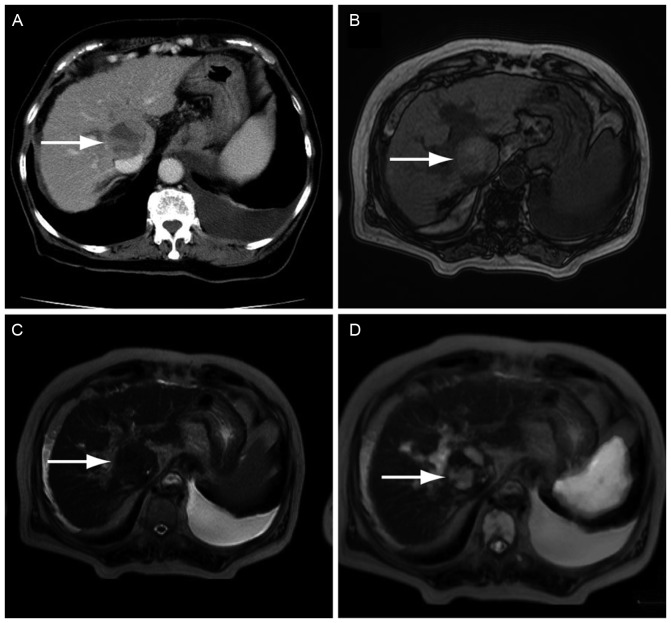
Figure 1.
Imaging examination in the representative case of a 73-year-old patient diagnosed with hepatocellular carcinoma. The tumor was successfully detected with DWIBS/T2. (A) CECT scanning demonstrated a space-occupying lesion with mixed enhancement (arrow). (B) T1WI showed an unclear mass-like lesion. (C) Detection of the lesion was difficult in a T2WI when compared with the CECT scan. (D) DWIBS/T2 clearly showed a high signal. DWIBS/T2, diffusion-weighted whole-body imaging with background body signal suppression/T2-weighted image fusion; CECT, contrast-enhanced computed tomography; WI, weighted image.

