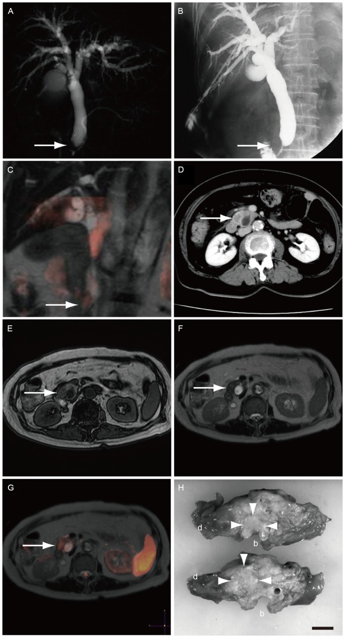
Figure 2.
Imaging examination in the representative case of a 75-year-old female patient diagnosed with pancreatic cancer. The tumor was successfully detected by DWIBS/T2. (A) Magnetic resonance cholangiopancreatography showed obstruction of the common bile duct near the papilla of Vater (arrow). (B) Percutaneous transhepatic biliary drainage was performed. (C) A coronal section of DWIBS/T2 demonstrated a high signal on the obstruction. (D) CECT revealed an irregularly shaped low-density area in the head of the pancreas. (E) T1WI and (F) T2WI scans did not show clear presence of pancreatic cancer. (G) A transverse section of DWIBS/T2 demonstrated a high signal in the head of the pancreas. (H) Surgical specimens confirmed the diagnosis of pancreatic cancer in the head of the pancreas close to common bile duct (circled with arrowheads). Scale bar, 1 cm; b, common bile duct; d, duodenum; DWIBS/T2, diffusion-weighted whole-body imaging with background body signal suppression/T2-weighted image fusion; CECT, contrast-enhanced computed tomography; WI, weighted image.

