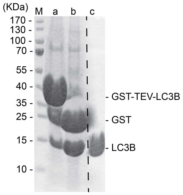
Fig. 3.
15% SDS-PAGE showing LC3B (1–120) at different steps of purification. Lane M shows the molecular weight markers. Lane a shows constituents of bead slurry after binding GS4B resin. Lane b shows constituents of bead slurry after thrombin treatment. Lane c shows final purified LC3B (1–120) after gel filtration chromatography.

