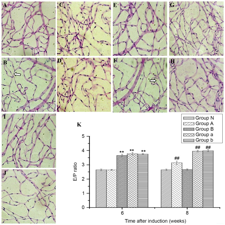
Figure 3.
Periodic acid-Schiff staining of retinal tissues in normal and diabetes mellitus rats to observe changes in the ratio of endotheliocytes to pericytes and whether there were acellular capillaries (magnification, ×400). Diabetic rats from group a at (A) six and (B) eight weeks; from group A at (C) six and (D) eight weeks; from group b at (E) six and (F) eight weeks; from group B at (G) six and (H) eight weeks; and from normal rats at (I) six and (J) eight weeks. (K) The ratio of endotheliocytes to pericytes at six and eight weeks after diabetes induction. White arrows indicate acellular capillaries. Bars indicate 95% confidence intervals. **P<0.01 vs. groups N and A at six weeks and ##P<0.01 vs. groups N and B at eight weeks. E/P, respectively, ratio of endotheliocytes to pericytes.

