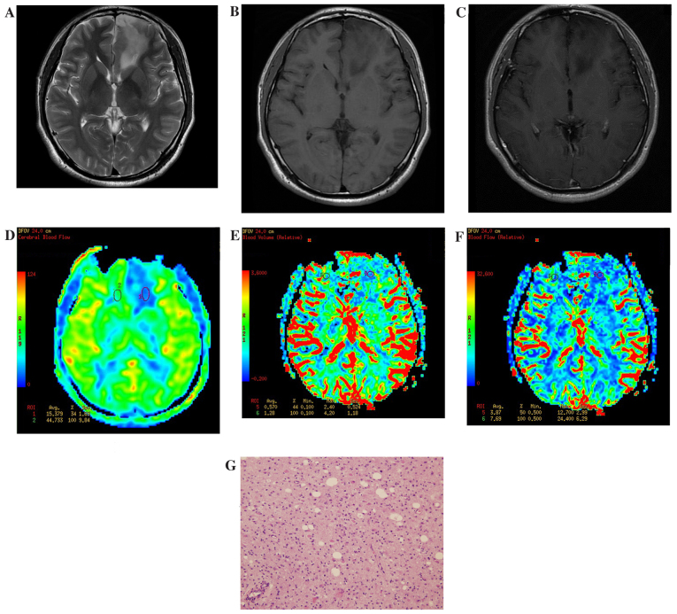
Figure 1.
Results of imaging of a patient with a low-grade glioma located at the left side of the frontal lobe. (A) Routine T2WI. The focal lesion showed a slightly higher signal. (B) Routine T1WI. The focal lesion showed a slightly low signal. (C) Enhanced T1WI. The focal lesion had no obvious enhancement. (D) ASL-CBF imaging. Low perfusion of the tumor was observed, with a ASL-rCBF value of 0.46. (E) DSC-CBV imaging. Low perfusion of the tumor was observed, with a DSC-rCBV value of 0.44. (F) DSC-CBF imaging. Low perfusion of the tumor was observed, with a DSC-rCBF value of 0.5. (G) Hematoxylin and eosin staining of a low-grade glioma specimen (magnification, ×40). The volume of tumor cells was large with abundant cytoplasm. WI, weighted imaging; ASL, arterial spin labelling; CBF, cerebral blood flow; DSC, dynamic susceptibility contrast; CBV, cerebral blood volume; r, relative.

