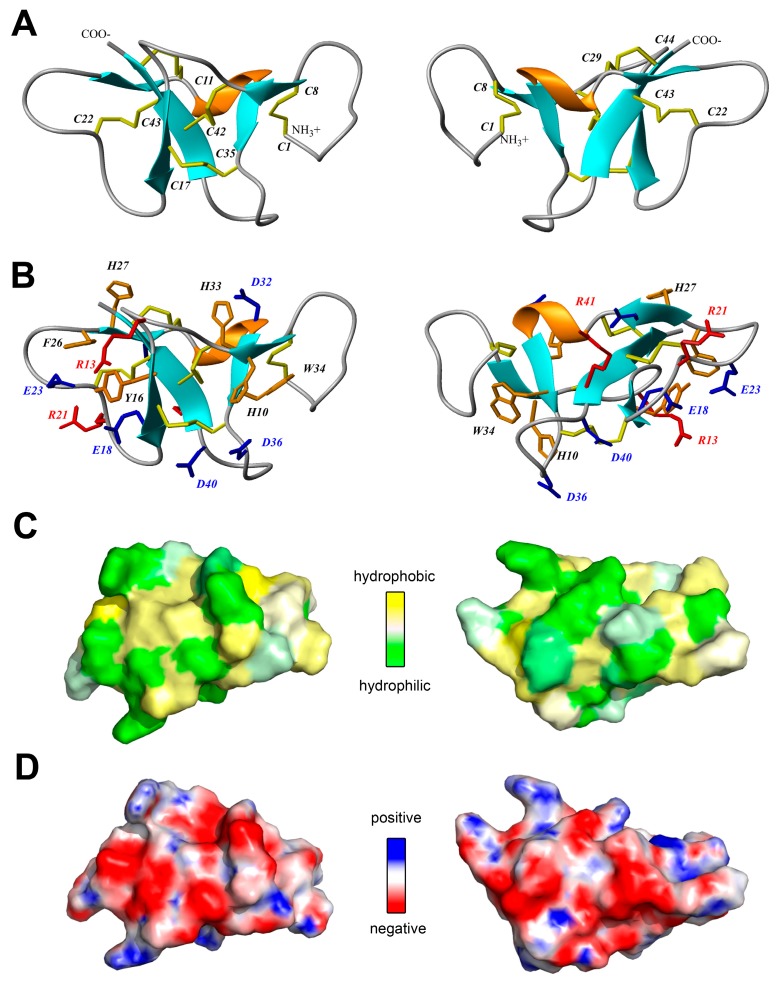
Figure 4.
Two-sided view of the determined spatial structure of Ueq 12-1 (PDB ID: 5LAH). (A,B) Ribbon representation. Disulfide bridges, positively and negatively charged side chains and aromatic residues are shown by yellow, red, blue and orange sticks, respectively; (C) Hydrophobicity of the Ueq 12-1 surface. Contact surface of Ueq 12-1 is colored from yellow (hydrophobic) to green (hydrophilic), according to the molecular hydrophobic potential (MHP) [30]; (D) molecular surface of Ueq 12-1 is painted according to the electrostatic potential.

