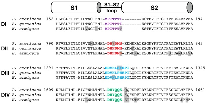
Figure 11.
Alignment of the S1–S2 regions from each of the four domains (DI–DIV) in the NaV channels from P. americana (UniProt D0E0C1), B. germanica (UniProt O01307), and H. armigera (deduced from the published genome). The S1–S2 extracellular loops from the four domains are coloured purple (DI), red (DII), blue (DIII) and green (DIV). Differences in amino acid sequence between species are highlighted in grey. The boundaries of S1 and S2 are based on the recently determined structure of the P. americana NaVPaS channel [39].

