
Figure 2.
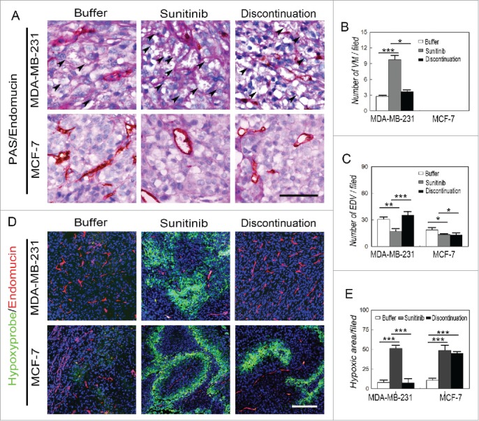
Effects of sunitinib treatment on the microcirculation patterns in TNBC tumors. (A) PAS and endomucin double staining in TNBCMDA-MB-231 and non-TNBC MCF-7 tumors. There were VM channels formed by PAS-positive molecules and tumor cells in the human TNBCMDA-MB-231 tumors, whereas no VM channels were observed in the human non-TNBC MCF-7 tumors. Growth of endothelium-dependent vessels in the TNBC MDA-MB-231 tumors was blocked by sunitinib, during which the number of VM channels significantly increased. Endothelium-dependent vessel growth rebounded in MDA-MB-231 tumors after treatment discontinuation, but there was no significant difference in the number of endothelium-dependent vessels between sunitinib-treated and post-treatment non-TNBC MCF-7 tumors. (B)Quantification of VM channels in the different groups. (C)Quantification of endothelium-dependent vessels in the different groups. (D)Hypoxia and endomucin double staining inTNBCMDA-MB-231 and non-TNBC MCF-7 tumors. There were more hypoxic regions in the sunitinib-treatedMDA-MB-231 tumors compared with those in the other groups when the endothelium-dependent vessels are inhibited. After treatment discontinuation, the hypoxic area in the MDA-MB-231 tumors decreased after the endothelium-dependent vessels rebounded. The hypoxic area in the MCF-7 tumors was similar in the sunitinib-treated and discontinued groups. (E)Quantification of the hypoxic area in the different groups. The scale bar indicates 100 μm, and the error bar indicates the standard deviation (SD). * P < 0.05, **P < 0.01, ***P < 0.001.