
Figure 4.
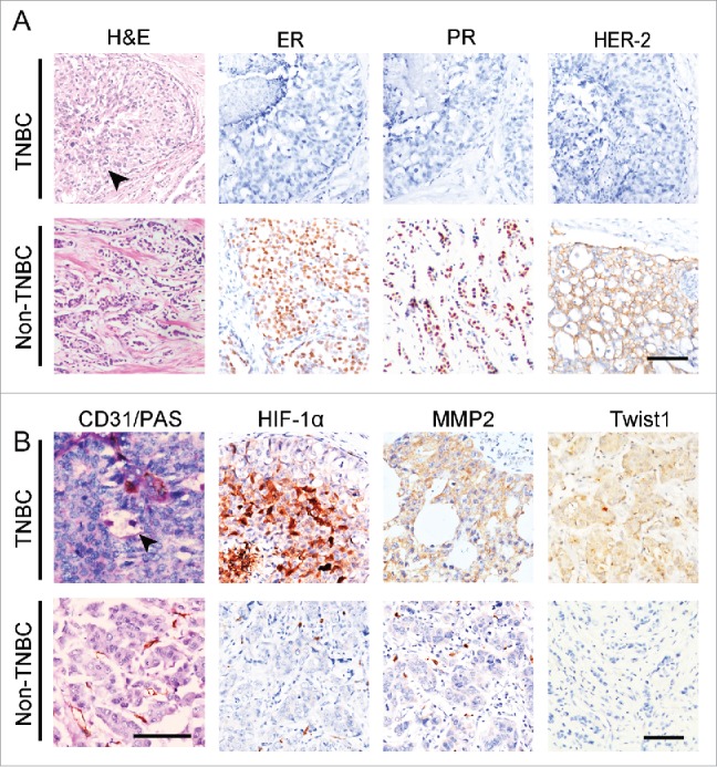
The expression of Twist1 in human TNBC and non-TNBC. (A) Morphological characteristics and genotypes of human TNBC and non-TNBC. H&E staining indicates that TNBC tumor nests comprise poorly differentiated small tumor cells, and there is necrosis in the center of a tumor nest (black arrow). Human TNBC tumors do not express ER, PR, orHER2. (B) PAS and CD31double staining showed that TNBC has more VM channels compared with non-TNBC. The arrow indicates a VM channel that is formed by PAS-positive matrix and tumor cells in TNBC. IHC staining indicates that TNBC tumors express higher levels of HIF-1α, MMP2 and Twist1 than non-TNBC tumors.