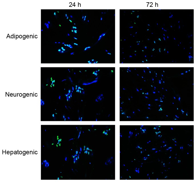
Figure 2.

Protein transduction and visualization in human fibroblasts 24 and 72 h after hemagglutinating virus of Japan envelope transduction of fibroblasts. Cells were stained with Hochest 33342 (blue) for nuclei and immunostained with anti-His antibody (green) for transcription factors (magnification, ×100).
