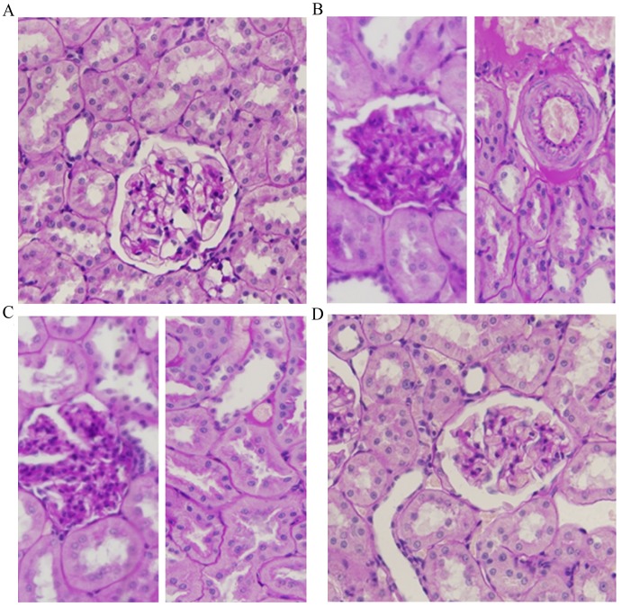
Figure 2.
Hematoxylin and eosin staining of kidney sections from diabetic rats with and without RSV treatment. (A) Normal glomerulus and tubules of an NG rat. (B) Glomerulus and tubules of a DM rat. Glomerular thickening, interstitial fibrosis and hyaline changes, epithelial cellular vacuolar degeneration, hyaline casts and arteriolopathy are visible. (C) Glomerulus and tubules of a DM rat treated with DMSO vehicle control. Histological alterations of glomeruli and arterioles are similar to those observed in the DM group. (D) Glomerulus and tubules of a DM rat treated with RSV. Milder histological alterations of glomeruli and arterioles are observed. Magnification, ×400. NG, normal control group; DM, diabetes mellitus control group; DM + DMSO, diabetic rats treated with vehicle; DM + RSV, diabetic rats treated with RSV. The dark blue color represents the nuclei and the pink color represents cytoplasm, fibronectin and red blood cells.

