Figure 1.
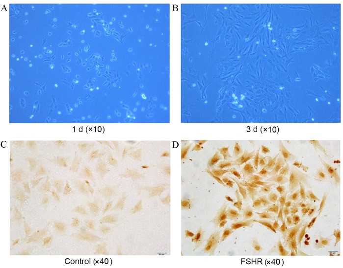
GC culture and characterization. Morphology (phase-contrast images; magnification, ×10) of rat ovary GCs in culture at (A) 1 d and (B) 3 d time points. Images of (C) control and (D) anti-FSHR antibody stained with cultured GCs. GCs with FSHR were stained brown. GC, granulosa cell; d, day(s); FSHR, follicle-stimulating hormone receptor.

