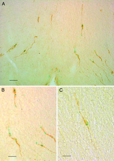
Fig. 1.
Localization of GPR54 to GnRH neurons. (A and B) Coronal sections from gpr54–/– mice preoptic hypothalami, showing neurons and dendrites double stained for β-galactosidase (blue) and GnRH (brown). (C) Representative medium-power field of preoptic areas. [Scale bars: 35 μm(A), 80 μm(B), and 45 μm(C).]

