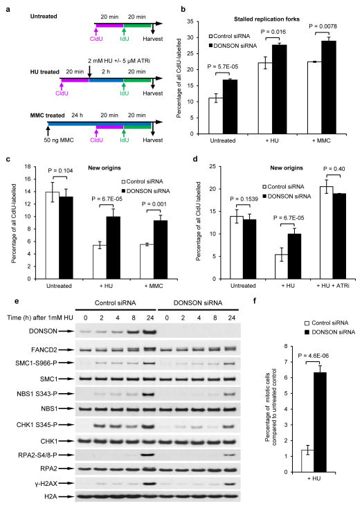
Figure 5. Depletion of DONSON compromises activation of cell cycle checkpoints.
(a–c) Loss of DONSON results in replication fork instability that is exacerbated by replication stress. (a) HeLa cells transfected with either control or DONSON siRNA were pulsed with CldU, exposed to 2 mM HU for 2 h, and then pulsed with IdU. Alternatively, cells were exposed to 50 ng/ml MMC for 24 h, and pulsed with sequential pulses of CldU and IdU (see schematic). DNA fibres were quantified, and the percentage of (b) stalled forks and (c) new origins are displayed (in all cases n=3). (d) Loss of DONSON is epistatic with ATR inhibition. Replication fork analysis of HeLa cells transfected with either control or DONSON siRNA. Cells were pulsed with CldU, exposed to 2 mM HU +/− 5μM ATR inhibitor for 2 h, and then pulsed with IdU (n=3). New origins (2nd label origin) were counted as an indicator of intra-S phase checkpoint activation. (e) Cells lacking DONSON exhibit defective or delayed ATR activation in response to replication stress. Whole cell extracts of HeLa cells transfected with either control or DONSON siRNA were subjected to immunoblot analysis using the indicated antibodies following treatment with 1 mM HU (n=2). (f) The percentage of mitotic cells following exposure to 1 mM HU for 24 h (from (e)) was determined by flow cytometry, using antibodies to phosphorylated histone H3-Ser10 (a marker of mitosis) (n=5).

