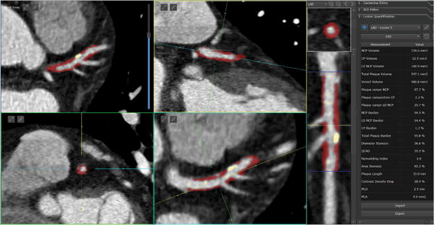
Figure 3.

Standardized quantification of high-risk plaque lesion in the left anterior descending artery. Full color available online. The quantification allows standardized measurement of several parameters such as maximal stenosis, volumes of non-calcified (red) and calcified (yellow) plaques, total plaque volume, plaque composition, length of the lesion and drop of CT contrast.
