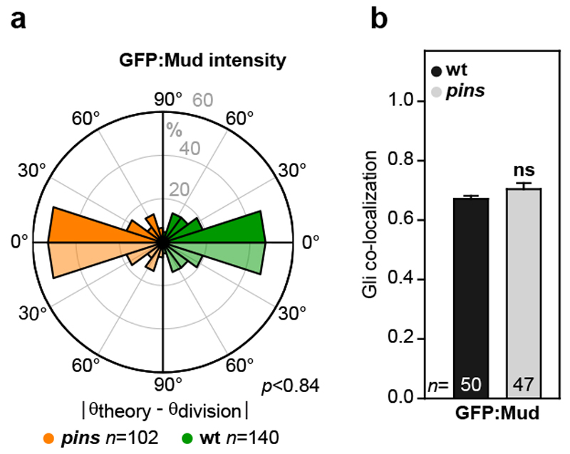
Extended Data Figure 7. Pins does not contribute to Mud-dependent epithelial cell division orientation.
(a) Rose plots of the difference between the theoretically predicted (θtheory) and the experimental division (θdivision) orientation of the mitotic spindle in pins tissue (orange left rose plot) and wt tissue (green right) based on the GFP:Mud intensity. To facilitate the comparison between the left and the right rose plots, the data are duplicated relative to 0° line (light orange and light green).Number of cells (n) analysed is indicated. p value, Kolmogorov-Smirnov test.
(b) Quantifications of the co-localization of GFP:Mud with Gli in pins in metaphase cells (±s.e.m.). Number of cells (n) analysed is indicated. ns: not significant (Student t-test).

