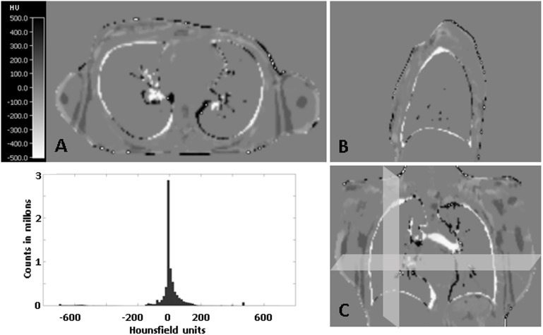
Fig 2.
Example of a subtraction image of the MR-based μ-map and the registered CT-based μ-map of a patient in which the LACs from the lung and the bone tissue were replaced by the LACs from the MR- μ-map in axial view (A), sagittal view (B) and coronal view (C). The histogram analysis of all patients shows that most of the voxels within the patient from the subtraction images provide HU of around 0 (>90% of the voxels show a deviation of more than 10% from the maximum value (0.1cm-1)) which stands for the high precision of the performed registration.

